


Preamble
Guidelines and Expert Consensus documents aim to present all the relevant evidence on a particular issue in order to help physicians to weigh the benefits and risks of a particular diagnostic or therapeutic procedure. They should be helpful in everyday clinical decision-making.
A great number of Guidelines and Expert Consensus Documents have been issued in recent years by the European Society of Cardiology (ESC) and by different organizations and other related societies. This profusion can put at stake the authority and validity of guidelines, which can only be guaranteed if they have been developed by an unquestionable decision-making process. This is one of the reasons why the ESC and others have issued recommendations for formulating and issuing Guidelines and Expert Consensus Documents.
In spite of the fact that standards for issuing good quality Guidelines and Expert Consensus Documents are well defined, recent surveys of Guidelines and Expert Consensus Documents published in peer-reviewed journals between 1985 and 1998 have shown that methodological standards were not complied with in the vast majority of cases. It is therefore of great importance that guidelines and recommendations are presented in formats that are easily interpreted. Subsequently, their implementation programmes must also be well conducted. Attempts have been made to determine whether guidelines improve the quality of clinical practice and the utilization of health resources.
The ESC Committee for Practice Guidelines (CPG) supervises and coordinates the preparation of new Guidelines and Expert Consensus Documents produced by Task Forces, expert groups or consensus panels. The chosen experts in these writing panels are asked to provide disclosure statements of all relationships they may have which might be perceived as real or potential conflicts of interest. These disclosure forms are kept on file at the European Heart House, headquarters of the ESC. The Committee is also responsible for the endorsement of these Guidelines and Expert Consensus Documents or statements.
The Task Force has classified and ranked the usefulness or efficacy of the recommended procedure and/or treatments and the Level of Evidence as indicated in the tables below:
Classes of Recommendations
| Class I | Evidence and/or general agreement that a given diagnostic procedure/treatment is beneficial, useful and effective; |
| Class II | Conflicting evidence and/or a divergence of opinion about the usefulness/efficacy of the treatment; |
| Class IIa | Weight of evidence/opinion is in favour of usefulness/efficacy; |
| Class IIb | Usefulness/efficacy is less well established by evidence/opinion; |
| Class III* | Evidence or general agreement that the treatment is not useful/effective and in some cases may be harmful. |
*Use of Class III is discouraged by the ESC.
Levels of Evidence
| Level of Evidence A | Data derived from multiple randomized clinical trials or meta-analyses |
| Level of Evidence B | Data derived from a single randomized clinical trial or large non-randomized studies |
| Level of Evidence C | Consensus of opinion of the experts and/or small studies; retrospective studies and registries |
1 Introduction
The aim of these guidelines is to describe the rationale behind the diagnosis and treatment of acute heart failure (AHF) in the adult population.
The Committee for Practice Guidelines (CPG) of the European Society of Cardiology nominated the Task Force for the AHF Guidelines. The Task Force included representatives from the Heart Failure Association of the ESC and members of the European Society of Intensive Care Medicine (ESICM). The Task Force recommendations were circulated among a review board and approved by the CPG of the ESC and by the ESICM. Together with the Guidelines for the diagnosis and treatment of chronic heart failure1 these Guidelines form the recommendations on diagnosis and treatment of heart failure.
The recommendations are also published as an unabridged version of the document,2 as a pocket guideline, and as an ESC educational product CD.
2 Epidemiology, aetiology, and clinical context
The combination of the aging of the population in many countries, and improved survival after acute myocardial infarction (AMI)3 has created a rapid growth in the number of patients currently living with chronic heart failure (CHF),4 with a concomitant increase in the number of hospitalizations for decompensated heart failure. Coronary heart disease is the aetiology of AHF in 60–70% of patients,5–7 particularly in the elderly population. In younger subjects, AHF is frequently caused by dilated cadiomyopathy, arrhythmia, congenital or valvular heart disease, or myocarditis. The causes and complications of AHF are described in Table 1.
The management of heart failure consumes 1–2% of health care expenditure in European countries,8,9 with around 75% relating to inpatient care. Advanced heart failure and related acute decompensation have become the single most costly medical syndrome in cardiology.10,11
Patients with AHF have a very poor prognosis. Mortality is particularly high in patients with acute myocardial infarction (AMI) accompanied by severe heart failure, with a 30% 12 month mortality.12 Likewise, in acute pulmonary oedema a 12% in-hospital and 40% 1 year mortality have been reported.13
About 45% of patients hospitalized with AHF will be rehospitalized at least once (and 15% at least twice) within twelve months.14,15 Estimates of the risk of death or rehospitalization within 60 days of admission vary from 30 to 60%, depending on the population studied.5,6,16–19
I Definitions, diagnostic steps, instrumentation and monitoring of the patient with AHF
3 Definition and clinical classification of AHF
3.1 Definition
Acute heart failure is defined as the rapid onset of symptoms and signs secondary to abnormal cardiac function. It may occur with or without previous cardiac disease. The cardiac dysfunction can be related to systolic or diastolic dysfunction, to abnormalities in cardiac rhythm, or to pre-load and after-load mismatch. It is often life threatening and requires urgent treatment.
AHF can present itself as acute de novo (new onset of acute heart failure in a patient without previously known cardiac dysfunction) or acute decompensation of chronic heart failure.
The patient with acute heart failure may present with one of several distinct clinical conditions (Table 2):
Acute decompensated heart failure (de novo or as decompensation of chronic heart failure) with signs and symptoms of acute heart failure, which are mild and do not fulfil criteria for cardiogenic shock, pulmonary oedema or hypertensive crisis.
Hypertensive AHF: Signs and symptoms of heart failure are accompanied by high blood pressure and relatively preserved left ventricular function with a chest radiograph compatible with acute pulmonary oedema.
Pulmonary oedema (verified by chest X-ray) accompanied by severe respiratory distress, with crackles over the lung and orthopnoea, with O2 saturation usually <90% on room air prior to treatment.
Cardiogenic shock: Cardiogenic shock is defined as evidence of tissue hypoperfusion induced by heart failure after correction of pre-load. There is no clear definition for haemodynamic parameters, which explains the differences in prevalence and outcome reported in studies (Table 2), but cardiogenic shock is usually characterized by reduced blood pressure (systolic BP <90 mmHg or a drop of mean arterial pressure>30 mmHg) and/or low urine output (<0.5 ml/kg/h), with a pulse rate >60 b.p.m. with or without evidence of organ congestion. There is a continuum from low cardiac output syndrome to cardiogenic shock.
High output failure is characterized by high cardiac output, usually with high heart rate (caused by arrhythmias, thyrotoxicosis, anaemia, Paget's disease, iatrogenic or by other mechanisms), with warm peripheries, pulmonary congestion, and sometimes with low BP as in septic shock.
Right heart failure is characterized by low output syndrome with increased jugular venous pressure, increased liver size and hypotension.
Various other classifications of the acute heart failure syndrome are utilized in coronary care and intensive care units. The Killip classification is based on clinical signs and chest X-ray findings, and the Forrester classification is based on clinical signs and haemodynamic characteristics. These classifications have been validated in acute heart failure after AMI and thus are best applied to acute de novo heart failure. The third ‘clinical severity’ classification has been validated in a cardiomyopathy service20 and is based on clinical findings.21 It is most applicable to chronic decompensated heart failure.22
3.1.1 Killip classification.
The Killip classification was designed to provide a clinical estimate of the severity of myocardial derangement in the treatment of AMI:23
StageI—No heart failure. No clinical signs of cardiac decompensation;
StageII—Heart failure. Diagnostic criteria include rales, S3 gallop and pulmonary venous hypertension. Pulmonary congestion with wet rales in the lower half of the lung fields;
StageIII—Severe heart failure. Frank pulmonary oedema with rales throughout the lung fields;
StageIV—Cardiogenic shock. Signs include hypotension (SBP≤90mmHg), and evidence of peripheral vasoconstriction such as oliguria, cyanosis and diaphoresis.
3.1.2 Forrester classification.
The Forrester AHF classification was also developed in AMI patients, and describes four groups according to clinical and haemodynamic status24 (Figure 1). Patients are classified clinically on the basis of peripheral hypoperfusion (filliform pulse, cold clammy skin, peripheral cyanosis, hypotension, tachycardia, confusion, oliguria) and pulmonary congestion (rales, abnormal chest X-ray), and haemodynamically on the basis of a depressed cardiac index (≤2.2 L/min/m2) and elevated pulmonary capillary pressure (>18 mmHg). The original paper defined the treatment strategy according to the clinical and haemodynamic status. Mortality was 2.2% in group I, 10.1% in group II, 22.4% in group III, and 55.5% in group IV.
3.1.3 ‘Clinical severity’ classification.
The clinical severity classification is based on observation of the peripheral circulation (perfusion) and on auscultation of the lungs (congestion). The patients can be classified as Class I (Group A) (warm and dry), Class II (Group B) (warm and wet), Class III (Group L) (cold and dry), and Class IV (Group C) (cold and wet). This classification has been validated prognostically in a cardiomyopathy service,20 and is therefore applicable to patients with chronic heart failure, whether hospitalized or outpatients.
3.2 The clinical syndrome of AHF
AHF is a clinical syndrome, with reduced cardiac output, tissue hypoperfusion, increase in the pulmonary capillary wedge pressure (PCWP), and tissue congestion. The underlying mechanism may be cardiac or extra-cardiac, and may be transient and reversible with resolution of the acute syndrome, or may induce permanent damage leading to chronic heart failure. The cardiac dysfunction can be related to systolic or diastolic myocardial dysfunction (mainly induced by ischaemia or infection), acute valvular dysfunction, pericardial tamponade, abnormalities of cardiac rhythm, or pre-load/after-load mismatch. Multiple extra-cardiac pathologies may result in acute heart failure by changing the cardiac loading conditions for example (i) increased after-load due to systemic or pulmonary hypertension or massive pulmonary emboli, (ii) increased pre-load due to increased volume intake or reduced excretion due to renal failure or endocrinopathy, or (iii) high output state due to infection, thyrotoxicosis, anaemia, Paget's disease. Heart failure can be complicated by co-existing end-organ disease. Severe heart failure can also induce multi-organ failure, which may be lethal.
Appropriate long-term medical therapy and, if possible, anatomical correction of the underlying pathology may prevent further AHF syndrome ‘attacks’ and improve the poor long-term prognosis associated with this syndrome.
The clinical AHF syndrome may be classified as predominantly left or right forward failure, left or right backward failure, or a combination of these.
3.2.1 Forward (left and right) AHF.
Forward acute heart failure may be mild-to-moderate with only effort fatigue, up to severe with manifestations of reduced tissue perfusion at rest with weakness, confusion, drowsiness, paleness with peripheral cyanosis, cold clammy skin, low blood pressure, filliform pulse, and oliguria, culminating in the full blown presentation of cardiogenic shock.
This syndrome may be induced by a large variety of pathologies. An adequate history may indicate the main diagnosis for example (i) acute coronary syndrome with the relevant risk factors, past history, and suggestive symptoms; (ii) acute myocarditis with a recent history suggestive of acute viral infection; (iii) acute valvular dysfunction with a history of chronic valve disease or valve surgery, infection with the possibility of bacterial endocarditis, or chest trauma; (iv) pulmonary embolism with a relevant history and suggestive symptoms; or (v) pericardial tamponade.
Physical examination of the cardiovascular system may be indicative of the main diagnosis, for example by distended neck veins and paradoxical pulse (pericardial tamponade), muffled heart sounds related to myocardial systolic dysfunction, or the disappearance of artificial valve sounds or an appropriate murmur indicating a valvular problem.
In forward AHF immediate management should include supportive treatment to improve cardiac output and tissue oxygenation. This can be achieved with vasodilating agents, fluid replacement to achieve an optimal pre-load, short-term inotropic support and (sometimes) intra-aortic balloon counterpulsation.
3.2.2 Left-heart backward failure.
Left-heart backward failure may be related to left ventricular dysfunction with varying degrees of severity, from mild-to-moderate with only exertional dyspnoea, to pulmonary oedema presenting with shortness of breath (dry cough, sometimes with frothy sputum), pallor or even cyanosis, cold clammy skin, and normal or elevated blood pressure. Fine rales are usually audible over the lung fields. Chest X-ray shows pulmonary congestion/oedema.
Pathology of the left heart may be responsible for this syndrome, including: myocardial dysfunction related to chronic existing conditions; acute insult such as myocardial ischaemia or infarction; aortic and mitral valve dysfunction; cardiac rhythm disturbances; or tumours of the left heart. Extra-cardiac pathologies may include severe hypertension, high output states (anaemia, thyrotoxicosis) and neurogenic states (brain tumours or trauma).
Physical examination of the cardiovascular system, including the apex beat, the quality of the heart sounds, the presence of murmurs, and auscultation of the lungs for fine rales and expiratory wheezing (‘cardiac asthma’) may be indicative of the main diagnosis.
In left heart backward failure patients should be treated mainly with vasodilation and the addition of diuretics, bronchodilators and narcotics, as required. Respiratory support may be necessary. This can either be with continuous positive airway pressure (CPAP) or non-invasive positive pressure ventilation, or in some circumstances invasive ventilation may be required following endotracheal intubation.
3.2.3 Right-heart backward failure.
The syndrome of acute right heart failure is related to pulmonary and right heart dysfunction, including exacerbations of chronic lung disease with pulmonary hypertension, or acute massive lung disease (e.g. massive pneumonia or pulmonary embolism), acute right ventricular infarction, tricuspid valve malfunction (traumatic or infectious), and acute or sub-acute pericardial disease. Advanced left heart disease progressing to right-sided failure should also be considered, and similarly long-standing congenital heart disease with evolving right ventricular failure should be taken into account. Non-cardiopulmonary pathologies include nephritic/nephrotic syndrome and end-stage liver disease. Various vasoactive peptide-secreting tumours should also be considered.
The typical presentation is with fatigue, pitting ankle oedema, tenderness in the upper abdomen (due to liver congestion), shortness of breath (with pleural effusion) and distension of the abdomen (with ascites). The full-blown syndrome includes anasarca with liver dysfunction and oliguria.
History and physical examination should confirm the syndrome of acute right heart failure, indicate the suspected diagnosis and guide further investigation, which is likely to include ECG, blood gases, D-dimer, chest X-ray, cardiac Doppler-echocardiography, angiography or chest CT scan.
In right heart backward failure fluid overload is managed with diuretics, including spironolactone, and sometimes with a short course of low dose (‘diuretic dose’) of dopamine. Concomitant treatment may include: antibiotics for pulmonary infection and bacterial endocarditis; Ca++ channel blockers, nitric oxide, or prostaglandins for primary pulmonary hypertension; and anticoagulants, thrombolytics, or thrombectomy for acute pulmonary embolism.
4 Pathophysiology of AHF
4.1 The vicious circle in the acute failing heart
The final common denominator in the syndrome of AHF is a critical inability of the myocardium to maintain a cardiac output sufficient to meet the demands of the peripheral circulation. Irrespective of the underlying cause of AHF, a vicious circle is activated that, if not appropriately treated, leads to chronic heart failure and death. This is shown in Figure 2, and is described in detail elsewhere.2
In order for patients with AHF to respond to treatment the myocardial dysfunction must be reversible. This is particularly important in AHF due to ischaemia, stunning or hibernation, where a dysfunctional myocardium can return to normal when appropriately treated.
4.2 Myocardial stunning
Myocardial stunning is the myocardial dysfunction that occurs following prolonged ischaemia, which may persist in the short-term even when normal blood flow is restored.25,26 The intensity and duration of stunning is dependent on the severity and duration of the preceding ischaemic insult.26
4.3 Hibernation
Hibernation is defined as an impairment of myocardial function due to severely reduced coronary blood flow although myocardial cells are still intact. By improving blood flow and oxygenation, hibernating myocardium can restore its normal function.27
Hibernating myocardium and stunning can co-exist. Hibernation improves in time with reinstitution of blood flow and oxygenation, whilst stunned myocardium retains inotropic reserve and can respond to inotropic stimulation.26,28 Since these mechanisms depend on the duration of myocardial damage, a rapid restoration of oxygenation and blood flow is mandatory to reverse these pathophysiological alterations.
5 Diagnosis of AHF
The diagnosis of AHF is based on the symptoms and clinical findings, supported by appropriate investigations such as ECG, chest X-ray, biomarkers, and Doppler-echocardiography (Figure 3). The patient should be classified according to previously described criteria for systolic and/or diastolic dysfunction (Figure 4), and by the characteristics of forward or backward left or right heart failure.
5.1 Clinical evaluation
Systematic clinical assessment of the peripheral circulation, venous filling, and peripheral temperature are important.
Right ventricular (RV) filling in decompensated heart failure may usually be evaluated from the central jugular venous pressure. When the internal jugular veins are impractical for evaluation (e.g. due to venous valves) the external jugular veins can be used. Caution is necessary in the interpretation of high measured central venous pressure (CVP) in AHF, as this may be a reflection of decreased venous compliance together with decreased RV compliance even in the presence of low RV filling.
Left sided filling pressure is assessed by chest auscultation, with the presence of wet rales in the lung fields usually indicating raised pressure. The confirmation, classification of severity, and clinical follow-up of pulmonary congestion and pleural effusions should be done using the chest X-ray.
Class I recommendation, level of evidence C
Again, in acute conditions the clinical evaluation of left-sided filling pressure may be misleading due to the rapidly evolving clinical situation. Cardiac palpation and auscultation for ventricular and atrial gallop rhythms (S3, S4) should be performed. The quality of the heart sounds, and the presence of atrial and ventricular gallops and valvular murmurs are important for diagnosis and clinical assessment. Assessment of the extent of arteriosclerosis by detecting missing pulses and the presence of carotid and abdominal bruits is often important, particularly in elderly subjects.
5.2 Electrocardiogram (ECG)
A normal ECG is uncommon in acute heart failure. The ECG is able to identify the rhythm, and may help determine the aetiology of AHF and assess the loading conditions of the heart. It is essential in the assessment of acute coronary syndromes.29–31 The ECG may also indicate acute right or left ventricular or atrial strain, perimyocarditis and pre-existing conditions such as left and right ventricular hypertrophy or dilated cardiomyopathy. Cardiac arrhythmia should be assessed in the 12-lead ECG as well as in continuous ECG monitoring.
5.3 Chest X-ray and imaging techniques
Chest X-ray and other imaging should be performed early for all patients with AHF to evaluate pre-existing chest or cardiac conditions (cardiac size and shape) and to assess pulmonary congestion. It is used both for confirmation of the diagnosis, and for follow-up of improvement or unsatisfactory response to therapy. Chest X-ray allows the differential diagnosis of left heart failure from inflammatory or infectious lung diseases. Chest CT scan with or without contrast angiography and scintigraphy may be used to clarify the pulmonary pathology and diagnose major pulmonary embolism. CT scan or transesophageal echocardiography should be used in cases of suspicion of aortic dissection.
5.4 Laboratory tests
A number of laboratory tests should be performed in AHF patients (Table 3). Arterial blood gas analysis (Astrup) enables assessment of oxygenation (pO2), respiratory adequacy (pCO2), acid–base balance (pH), and base deficit, and should be performed in all patients with severe heart failure. Non-invasive measurement with pulse oximetry and end-tidal CO2 can often replace Astrup (Level of evidence C) but not in very low output, vasocontricted shock states. Measurement of venous O2 saturation (i.e. in the jugular vein) may be useful for an estimation of the total body oxygen supply-demand balance.
Plasma B-type natriuretic peptide (BNP) is released from the cardiac ventricles in response to increased wall stretch and volume overload and has been used to exclude and/or identify congestive heart failure (CHF) in patients admitted, for dyspnoea, to the emergency department.1,32 Decision cut points of 300 pg/mL for NT-proBNP and 100 pg/mL for BNP have been proposed, but the older population has been poorly studied. During ‘flash’ pulmonary oedema, BNP levels may remain normal at the time of admission. Otherwise, BNP has a good negative predictive value to exclude heart failure.33 Various clinical conditions may affect the BNP concentration including renal failure and septicaemia. If elevated concentrations are present, further diagnostic tests are required. If AHF is confirmed, increased levels of plasma BNP and NT-pro BNP carry important prognostic information. The exact role of BNP remains to be fully clarified.34
5.5 Echocardiography
Echocardiography is an essential tool for the evaluation of the functional and structural changes underlying or associated with AHF, as well as in the assessment of acute coronary syndromes.
Class I recommendation, level of evidence C
Echocardiography with Doppler imaging should be used to evaluate and monitor regional and global left and right ventricular function, valvular structure and function, possible pericardial pathology, mechanical complications of acute myocardial infarction and—on rare occasions—space occupying lesions. Cardiac output can be estimated by appropriate Doppler aortic or pulmonary time velocity contour measurements. An appropriate echo-Doppler study can also estimate pulmonary artery pressures (from the tricuspid regurgitation jet) and has also been used for the monitoring of left ventricular pre-load.35–37 Echocardiography has not been validated with right heart catheterization in patients with AHF.38
5.6 Other investigations
In cases of coronary-artery-related complications such as unstable angina or myocardial infarction, angiography is important and angiography-based revascularization therapy has been shown to improve prognosis.29,30
Class I recommendation, level of evidence B
Coronary arteriography is also often indicated in prolonged AHF, unexplained by other investigations, as recommended in the guidelines for diagnosis of CHF.1
Insertion of a pulmonary artery catheter (PAC) may assist in making the diagnosis of AHF. See Section 7.2.3 for further details.
6 Goals of the treatment of AHF
The immediate goals are to improve symptoms and to stabilize the haemodynamic condition (Table 4, Figure 5).40–51 An improvement in haemodynamic parameters only may be misleading, however, and a concomitant improvement in symptoms (dyspnoea and/or fatigue) is generally required.52 These short-term benefits must also be accompanied by favourable effects on longer-term outcomes. This is likely to be achieved by avoidance, or limitation, of myocardial damage.
Another objective of treatment is reduction in the clinical signs of HF. A reduction in body weight, and/or an increase in diuresis, are beneficial effects of therapy in congestive and oliguric patients with AHF.44,53 Similarly, an improvement in oxygen saturation, renal and/or hepatic function, and/or serum electrolytes are meaningful goals of treatment. Plasma BNP concentration can reflect haemodynamic improvement and decreased levels are beneficial.
Beneficial effects of therapy on outcome include reductions in the duration of intravenous vasoactive therapy, the length of stay, and the readmission rate with an increase in the time to readmission.52,54,55 A reduction in both in-hospital and long-term mortality is also a major goal of treatment.
Lastly, a favourable safety and tolerability profile is also necessary for any treatment used in patients with AHF. Any agent used in this condition should be associated with a low withdrawal rate with a relatively low incidence of untoward side effects.
6.1 Organization of the treatment of AHF
Best results are achieved if patients with AHF are treated promptly by expert staff in areas reserved for heart failure patients. An experienced cardiologist and/or other suitably trained staff should treat AHF patients. The diagnostic services should provide early access to diagnostic procedures such as echocardiography and coronary angiography, as needed.
Treatment of patients with AHF requires a treatment plan in the hospital system.50
Class I recommendation, level of evidence B
Comparative studies have shown shorter hospitalization time in patients treated by staff trained in heart failure management.17 The treatment of AHF should be followed by a subsequent HF clinic programme when applicable and as recommended by ESC guidelines.1
The care and information needs of the acutely ill patient and his/her family will usually be addressed by expert nurses.
Heart failure staff nurses and cardiology/heart failure/intensive care specialists should be given the opportunity for continuing professional education.
Recommendations on the standard structure, nursing staff and equipment requirements in intensive cardiology care units and relevant step-down care units based on the expert opinion of the Working Group of Acute Cardiac Care are under preparation.
7 Instrumentation and monitoring of patients in AHF
Monitoring of the patient with AHF should be initiated as soon as possible after his/her arrival at the emergency unit, concurrently with ongoing diagnostic measures addressed at determining the primary aetiology. The types and level of monitoring required for any individual patient vary widely depending on the severity of the cardiac decompensation and the response to initial therapy. Local logistic issues may also be relevant. The guidelines on monitoring discussed here are based on expert opinion.
7.1 Non-invasive monitoring
In all critically ill patients, BP measurements should be made routinely; blood pressure, temperature, respiratory rate, heart rate, the electrocardiogram and blood pressure is mandatory. Some laboratory tests should be done repeatedly i.e. electrolytes, creatinine and glucose or markers for infection or other metabolic disorders. Hypo– or hyperkalaemia must be controlled. These can all be monitored easily and accurately with modern automated equipment. If the patient becomes more unwell, the frequency of these observations will need to be increased.
ECG monitoring (arrhythmias and ST segment) is necessary during the acute decompensation phase, particularly if ischaemia or arrhythmia is responsible for the acute event.
Class I recommendation, level of evidence C
Maintenance of normal blood pressure is critical during the initiation of therapy, and consequently it should be measured regularly (e.g. every 5 minutes), until the dosage of vasodilators, diuretics or inotropes has been stabilized. The reliability of non-invasive, automatic plethysmographic measurement of blood pressure is good in the absence of intense vasoconstriction and very high heart rate.
Class I recommendation, level of evidence C
The pulse oximeter is a simple non-invasive device that estimates the arterial saturation of haemoglobin with oxygen (SaO2). The estimate of the SaO2 is usually within 2% of a measured value from a co-oximeter, unless the patient is in cardiogenic shock. The pulse oximeter should be used continuously on any unstable patient who is being treated with a fraction of inspired oxygen (FiO2) that is greater than in air. It should also be used at regular intervals (every hour) on any patient receiving oxygen therapy for an acute decompensation.
Class I recommendation, level of evidence C
Cardiac output and pre-load can be monitored non-invasively with the use of Doppler techniques (see Section 5.5.). There is little to no evidence to help choose which of these to monitor and it makes no difference as long as the limitations of individual monitoring devices are understood and the data are used appropriately.
Class IIb recommendation, level of evidence C
7.2 Invasive monitoring
7.2.1 Arterial line.
The indications for the insertion of an in-dwelling arterial catheter are the need for either continuous beat-to-beat analysis of arterial blood pressure due to haemodynamic instability or the requirement for multiple arterial blood analyses. The complication rate for the insertion of a 20-gauge 2-inch radial artery catheter is low.
Class IIb recommendation, level of evidence C
7.2.2 Central venous pressure lines.
Central venous lines provide access to the central venous circulation and are therefore useful for the delivery of fluids and drugs and can also be used to monitor the CVP and venous oxygen saturation (SvO2) in the superior vena cava (SVC) or right atrium, which provides an estimate of oxygen transport.
Class II b recommendation, level of evidence C
Caution has to be advised, however, to avoid the over-interpretation of right atrial pressure measurements, as these rarely correlate with left atrial pressures, and therefore left ventricular (LV) filling pressures, in patients with AHF. CVP measurements are also affected by the presence of significant tricuspid regurgitation and positive end-expiratory pressure (PEEP) ventilation.
Class I recommendation, level of evidence C
7.2.3 Pulmonary artery catheter.
The pulmonary artery catheter (PAC) is a balloon flotation catheter that measures pressures in the superior vena cava (SVC), right atrium, right ventricle and pulmonary artery as well as cardiac output. Modern catheters can measure the cardiac output semi-continuously as well as the mixed venous oxygen saturation and right ventricular end diastolic volume and ejection fraction.
Although the insertion of a PAC for the diagnosis of AHF is usually unnecessary, it can be used to distinguish between a cardiogenic and a non-cardiogenic mechanism in complex patients with concurrent cardiac and pulmonary disease. The PAC is also frequently used to estimate PCWP, cardiac output and other haemodynamic variables and therefore guide therapy in the presence of severe diffuse pulmonary pathology or ongoing haemodynamic compromise not resolved by initial therapy.57,58 PCWP is not an accurate reflection of left ventricular end–diastolic pressure (LVEDP) in patients with mitral stenosis (MS) aortic regurgitation (AR), ventricular interdependence, high airway pressure, or stiff LV, due to, for example, left ventricular hypertrophy (LVH), diabetes, fibrosis, inotropes, obesity, ischaemia. Severe tricuspid regurgitation, frequently found in patients with AHF, can overestimate or underestimate cardiac output measured by thermodilution.
Several retrospective studies assessing the use of the PAC in acute myocardial infarction demonstrated increased mortality with the PAC. These observations were partially explained by case-mix differences between the groups of the study.59–61 Similar observational findings have subsequently been reported in other groups of patients.47,61,62 A recent prospective randomized study enrolling a mixed group of critically ill patients failed to demonstrate a difference in outcome, although randomization to the PAC led to increased fluid resuscitation within the first 24 h. The PAC did not cause harm to patients, rather it was the use of the information derived from the catheter (sometimes in an inappropriate fashion) that was detrimental.48
The use of a PAC is recommended in haemodynamically unstable patients who are not responding in a predictable fashion to traditional treatments, and in patients with a combination of congestion and hypoperfusion. In these cases it is inserted in order to ensure optimal fluid loading of the ventricles and to guide49 vasoactive therapies and inotropic agents (Table 5). Because the complications increase with the duration of its use, it is critical to insert the catheter when specific data are needed (usually regarding the fluid status of the patient) and to remove it as soon as it is of no further help (i.e. when diuretic and vasodilating therapy have been optimized).
Class IIb recommendation, level of evidence C
In cardiogenic shock and prolonged severe low output syndrome it is recommended that the mixed venous oxygen saturation from the pulmonary artery be measured as an estimation of oxygen extraction (SpO2–SvO2). The aim should be to maintain SvO2 above 65% in patients with AHF.
II Treatment of AHF
8 General medical issues in the treatment of AHF
Infections: Patients with advanced AHF are prone to infectious complications, commonly respiratory or urinary tract infections, septicaemia, or nosocomial infection with Gram positive bacteria. An increase in C-reactive protein (CRP) and a decrease in general condition may be the only signs of infection—fever may be absent. Meticulous infection control and measures to maintain skin integrity are mandatory. Routine cultures are recommended. Prompt antibiotic therapy should be given when indicated.
Diabetes: AHF is associated with impaired metabolic control. Hyperglycaemia occurs commonly. Routine hypoglycaemic drugs should be stopped and glycaemic control should be obtained by using short-acting insulin titrated according to repeated blood glucose measurements. Normoglycaemia improves survival in diabetic patients who are critically ill.50
Catabolic state: negative caloric and nitrogen balance is a problem in ongoing AHF. This is related to reduced caloric intake due to reduced intestinal absorption. Care should be undertaken to maintain calorie and nitrogen balance. Serum albumin concentration, as well as nitrogen balance, may help to monitor metabolic status.
Renal failure: a close interrelationship exists between AHF and renal failure. Both may cause, aggravate, and influence, the outcome of the other. Close monitoring of renal function is mandatory. Preservation of renal function is a major consideration in the selection of the appropriate therapeutic strategy for these patients.
9 Oxygen and ventilatory assistance
9.1 Rationale for using oxygen in AHF
The maintenance of an SaO2 within the normal range (95–98%) is important in order to maximize oxygen delivery to the tissues and tissue oxygenation, thus helping to prevent end-organ dysfunction and multiple organ failure.
Class I recommendation, level of evidence C
This is best achieved first by ensuring that there is a patent airway and then by administration of an increased FiO2. Endotracheal intubation is indicated if these measures fail to improve tissue oxygenation.
Class IIa recommendation, level of evidence C
Despite this intuitive approach to giving oxygen, there is little to no evidence available that giving increasing doses of oxygen results in an improved outcome. Studies have demonstrated that hyperoxia can be associated with reduced coronary blood flow, reduced cardiac output, increased blood pressure, increased systemic vascular resistance, and a trend to higher mortality.51
The administration of increased concentrations of oxygen to hypoxaemic patients with acute cardiac failure is unquestionably warranted.
Class IIa recommendation, level of evidence C
The use of increased concentrations of oxygen in patients without evidence of hypoxaemia is more controversial and may cause harm.63
9.2 Ventilatory support without endotracheal intubation (non-invasive ventilation)
Two techniques are used for ventilatory support: CPAP or non-invasive positive pressure ventilation (NIPPV). NIPPV is a method of providing mechanical ventilation to patients without the need for endotracheal intubation. There is a strong consensus that one of these two techniques should be used before endotracheal intubation and mechanical ventilation. Utilization of non-invasive techniques dramatically reduce the need for endotracheal intubation and mechanical ventilation.
9.2.1 Rationale.
Application of CPAP can cause pulmonary recruitment and is associated with an increase in the functional residual capacity. The improved pulmonary compliance, reduced transdiaphragmatic pressure swings, and decreased diaphragmatic activity can lead to a decrease in the overall work of breathing and therefore a decreased metabolic demand from the body. NIPPV is a more sophisticated technique that requires a ventilator. Addition of a PEEP to the inspiratory assistance results in a CPAP mode (also known as bilevel positive pressure support, BiPAP). The physiological benefits of this mode of ventilation are the same as for CPAP but also include the inspiratory assist which further reduces the work of breathing and the overall metabolic demand.
9.2.2 Evidence for the use of CPAP and NIPPV in left ventricular failure.
CPAP in patients with cardiogenic pulmonary oedema improves oxygenation, decreases symptoms and signs of AHF, and results in a decreased need for endotracheal intubation.64–68 The studies have been relatively small and therefore have not reported a statistically significant reduction in mortality. A systematic review69 following the first three trials suggested that CPAP was associated with a decreased need for intubation and a trend to decreased in-hospital mortality compared to standard therapy alone. Evidence was lacking, however, on the potential for CPAP to actually cause harm.
There have been three randomized controlled trials of the use of NIPPV in the setting of acute cardiogenic pulmonary oedema.70–72 NIPPV appears to decrease the need for endotracheal intubation, but this does not translate into a reduction in mortality or improvement in long-term function.
9.2.3 Conclusions.
The use of CPAP and NIPPV in acute cardiogenic pulmonary oedema is associated with a significant reduction in the need for tracheal intubation and mechanical ventilation.
Class IIa recommendation, level of evidence A
There are insufficient data to demonstrate a significant reduction in mortality; however, the data do not trend in that direction.
9.3 Mechanical ventilation with endotracheal intubation in AHF
Invasive mechanical ventilation (with endotracheal intubation) should not be used to reverse hypoxaemia that could be better restored by oxygen therapy, CPAP, or NIPPV, but rather to reverse AHF-induced respiratory muscle fatigue. The latter is the most frequent reason for endotracheal intubation and mechanical ventilation. Respiratory muscle fatigue may be diagnosed by a decrease in respiratory rate, associated with hypercapnia and confused state of mind.
Invasive mechanical ventilation should only be used if acute respiratory failure does not respond to vasodilators, oxygen therapy, and/or CPAP, or NIPPV. Another consideration should be the need for immediate intervention in a patient with pulmonary oedema secondary to ST-elevation acute coronary syndrome.
10 Medical treatment
10.1 Morphine and its analogues in AHF
Morphine is indicated in the early stage of the treatment of a patient admitted with severe AHF, especially if associated with restlessness and dyspnoea.
Class IIb recommendation, level of evidence B
Morphine induces venodilatation and mild arterial dilatation, and reduces heart rate.73 In most studies, iv boluses of morphine 3 mg were administered as soon as the intravenous line was inserted. This dosing can be repeated if required.
10.2 Anticoagulation
Anticoagulation is well established in acute coronary syndrome with or without heart failure.29 The same is true in atrial fibrillation.31 There is less evidence for the initiation of unfractionated heparin or low molecular weight heparin (LMWH) in AHF. A large placebo-controlled trial of enoxaparine 40 mg subcutaneoulsy in acutely ill and hospitalized patients, including a major group of heart failure patients, showed no clinical improvement but less venous thrombosis.74 There are no large comparative studies comparing LMWH to unfractionated heparin (given as 5000 IU twice or thrice daily). Careful monitoring of the coagulation system is mandatory in AHF as there is often concomitant liver dysfunction. LMWH is contraindicated if the creatinine clearance is below 30 mL/min or should be used with extreme care with monitoring of the anti-Factor Xa level.
10.3 Vasodilators in the treatment of AHF
Vasodilators are indicated in most patients with acute heart failure as first line therapy, if hypoperfusion is associated with an adequate blood pressure and signs of congestion with low diuresis, to open the peripheral circulation and to lower pre-load (Table 6).
10.3.1 Nitrates.
Nitrates relieve pulmonary congestion without compromising stroke volume or increasing myocardial oxygen demand in acute left heart failure, particularly in patients with acute coronary syndrome. At low doses they only induce venodilation, but as the dose is gradually increased they cause the arteries, including the coronary arteries, to dilate. With appropriate doses, nitrates exert balanced vasodilation of the venous and arterial sides of the circulation, thereby reducing LV pre-load and after-load, without impairing tissue perfusion. Their effect on cardiac output depends on pre-treatment pre-load and after-load and the ability of the heart to respond to baroreceptor-induced increases in sympathetic tone.
Initially nitrates may be given orally but intravenous nitrates are also well tolerated in AMI. Two randomized trials in AHF have established the efficacy of intravenous nitrates in combination with furosemide and have demonstrated that titration to the highest haemodynamically tolerable dose of nitrates with low dose furosemide is superior to high dose diuretic treatment alone.
Class I recommendation, level of evidence B
In one of these randomized studies furosemide and isosorbide dinitrate as bolus injections were tested and it was reported that intravenous high dose nitrate was more effective than furosemide treatment in controlling severe pulmonary oedema.75
In practical use nitrates have a U-shaped curve effect. If given in sub-optimal doses vasodilators may have a limited effect in preventing AHF recurrences. However, administration of high doses may also reduce their effectiveness. One disadvantage of nitrates is the rapid development of tolerance especially when given intravenously in high doses, limiting their effectiveness to 16–24 h only. Nitrates should be given at doses aimed at achieving optimal vasodilation, leading to an increase in cardiac index (CI) and decrease in pulmonary wedge pressure. Inappropriate vasodilation may induce a steep reduction in blood pressure, which may result in haemodynamic instability.
Nitroglycerin can be administered orally or by inhalation [glyceryl trinitrate (GTN) spray 400 µg (2 puffs) every 5–10 min], or buccally (isosorbide dinitrate 1 or 3 mg), while monitoring blood pressure. The intravenous administration and dosing of nitrates (glycerylnitrate 20 µg/min increasing dose to 200 µg/min, or isosorbide dinitrate 1–10 mg/h) should be done with extreme caution, under careful blood pressure monitoring, titrating the dose administered against blood pressure decrease. One should be particularly cautious when administering nitrates to a patient with aortic stenosis, although this therapy may help in these complex situations. The dose of nitrates should be reduced if systolic blood pressure falls below 90–100 mmHg and discontinued permanently if blood pressure drops further. From a practical point of view a reduction of 10 mmHg in mean arterial pressure should be achieved.
10.3.2 Sodium nitroprusside.
Sodium nitroprusside (SNP) (0.3 µg/kg/min up-titrating carefully to 1 µg/kg/min up to 5 µg/kg/min) is recommended in patients with severe heart failure, and in patients with predominantly increased after-load such as hypertensive heart failure or mitral regurgitation.
Class I recommendation, level of evidence C
SNP should be titrated cautiously and usually requires invasive arterial monitoring and close supervision. Prolonged administration may be associated with toxicity from its metabolites, thiocyanide and cyanide, and should be avoided especially in patients with severe renal or hepatic failure. Controlled trials with SNP in AHF are lacking and its administration in AMI has yielded equivocal results.76 SNP should be tapered down to avoid rebound effects. In AHF caused by acute coronary syndromes, nitrates are favoured over SNP as SNP may cause ‘coronary steal syndrome’.77,78
10.3.3 Nesiritide.
Recently, nesiritide, a new class of vasodilator, has been developed for the treatment of AHF.40 Nesiritide is a recombinant human brain or B-type natriuretic peptide (BNP) that is identical to the endogenous hormone. Nesiritide has venous, arterial, and coronary vasodilatory properties that reduce pre-load and after-load, and increase cardiac output without direct inotropic effects.
Systemic infusion of nesiritide in patients with CHF has beneficial haemodynamic actions, results in an enhanced sodium excretion, and suppression of the renin–angiotensin–aldosterone and sympathetic nervous systems.79 Nesiritide was compared to intravenous nitroglycerin and resulted in improvement in haemodynamics more effectively and with fewer adverse effects, although this did not translate into improvement in clinical outcome. Nesiritide may cause hypotension and some patients are non-responders.
10.3.4 Calcium antagonists.
Calcium antagonists are not recommended in the treatment of AHF. Diltiazem and verapamil, and dihydropyridines, should be considered contraindicated.
10.4 Angiotensin converting enzyme (ACE)-inhibitors in AHF
10.4.1 Indications.
ACE-inhibitors are not indicated in the early stabilization of patients with AHF.
Class IIb recommendation, level of evidence C
However, as these patients are at high risk, ACE-inhibitors have a role in early management of AHF patients and AMI. There is still debate on the selection of patients and the timing of initiation of ACE-inhibitor therapy.
10.4.2 Effects and mechanism of action.
The haemodynamic effects of ACE-inhibitors result from decreased formation of AII and increased levels of bradykinin, which in turn decreases total peripheral vascular resistances and promotes natriuresis. Short-term treatment is accompanied by a decrease in angiotensin II (AII) and aldosterone and an increase in angiotensin I and plasma renin activity.
There have been no efficacy studies of ACE-inhibitors in AHF to date. Studies with ACE-inhibitors in heart failure after myocardial infarction have focused on long-term effects.80,81 A recent meta-analysis found that mortality at 30 days was reduced from 7.6% in the placebo group to 7.1% in the ACE-inhibitor group [relative risk reduction 7% (95% CI 2–11%, P<0.004)]. This equates to about five fewer deaths per 1000 patients treated for 4–6 weeks [number needed to treat (NNT) to prevent one death=200]. The trials which selected high-risk patients found that ACE-inhibitors led to large relative and absolute reductions in mortality.82
10.4.3 Practical use.
Intravenous ACE-inhibition should be avoided. The initial dose of the ACE-inhibitor should be low and increased progressively after early stabilization within 48 h with monitoring of blood pressure and renal function. The duration of therapy, when initiated, should be at least six weeks.
Class I recommendation, level of evidence A
ACE-inhibitors should be used with caution in patients with marginal cardiac output as they may significantly reduce glomerular filtration.The risk of intolerance to the ACE-inhibitors is increased by the concomitant administration of non-steroid anti-inflammatory agents, and in the presence of bilateral renal artery stenosis.
10.5 Diuretics
10.5.1 Indications.
Administration of diuretics is indicated in patients with acute and acutely decompensated heart failure in the presence of symptoms secondary to fluid retention.
Class I recommendation, level of evidence B
The symptomatic benefits and their universal clinical acceptance have precluded a formal evaluation in large-scale randomized clinical trials.
10.5.2 Effects and mechanisms of action.
Diuretics increase the urine volume by enhancing the excretion of water, sodium chloride and other ions, leading to a decrease in plasma and extracellular fluid volume, total body water and sodium, a reduction in right and left ventricular filling pressures and a decrease in peripheral congestion and pulmonary oedema.83,84 Intravenous administration of loop diuretics also exerts a vasodilating effect, manifested by an early (5–30 min) decrease in right atrial and pulmonary wedge pressure as well as pulmonary resistances.85 With high bolus doses (>1 mg/kg) there is a risk of reflex vasoconstriction. As opposed to chronic use of diuretics, in severe decompensated heart failure the use of diuretics normalizes loading conditions and may reduce neurohormonal activation in the short term.86 Especially in acute coronary syndromes diuretics should be used in low doses and preference given to vasodilator therapy.87
10.5.3 Practical use.
Intravenous administration of loop diuretics (furosemide, bumetanide, torasemide), with a strong and brisk diuretic effect is the preferred choice in patients with AHF. Therapy can safely be initiated before hospital admission75,88–90 and the dose should be titrated according to the diuretic response and relief of congestive symptoms. Administration of a loading dose followed by continued infusion of furosemide or torasemide have been shown to be more effective than bolus alone.87,91–95 Thiazides96–98 and spironolactone99 can be used in association with loop diuretics, the combination in low doses being more effective and having with fewer secondary effects than the use of higher doses of a single drug.96–99 Combination of loop diuretics with dobutamine, dopamine92 or nitrates88 is also a therapeutic approach that is more effective and produces fewer secondary effects than increasing the dose of the diuretic.100
Class IIb recommendation, level of evidence C
Table 7 lists the recommendations for the practical use of diuretics. Table 8 gives the recommended doses of commonly used diuretics in heart failure.
10.5.4 Diuretic resistance.
Diuretic resistance is defined as the clinical state in which diuretic response is diminished or lost before the therapeutic goal of oedema relief has been achieved.101 Such resistance is associated with a poor prognosis.102 It is more frequent in patients with chronic, severe heart failure on long-term diuretic therapy, although it has also been reported with acute volume depletion after intravenous administration of loop diuretics.103 Diuretic resistance can be attributed to a number of factors (Table 9).101,103 A number of therapeutic approaches to overcome diuretic resistance have been explored (Table 10), and in clinical practice different strategies may be of value in a particular patient. Continuous infusion of furosemide is more effective than individual boluses.104
10.5.5 Secondary effects, drug interactions.
Although diuretics can be used safely in the majority of patients, secondary effects are frequent and may be life-threatening. They include neurohormonal activation, especially of the angiotensin–aldosterone system and the sympathetic nervous system97, hypokalaemia, hypomagnesaemia, and hypochloraemic alkalosis that may lead to severe arrhythmias92, and nephrotoxicity and aggravation of renal failure.100,105 Excessive diuresis may reduce venous pressure, pulmonary wedge pressure and diastolic filling excessively, leading to a reduction in stroke volume and cardiac output, particularly in patients with severe heart failure and predominant diastolic failure or ischaemic RV dysfunction. Intravenous administration of acetazolamide (1 or 2 doses) may be helpful for the correction of alkalosis.106
10.5.6 New diuretic agents.
Some new compounds with diuretic and other effects are under investigation, including vasopressin V2 receptor antagonists, brain natriuretic peptides (see Section 10.3.3) and adenosine receptor antagonists.
10.6 β-blocking agents
10.6.1 Indications and rationale for β-blocking agents.
There has been no study with β-blocker therapy in AHF targeted to acutely improve the condition. On the contrary, AHF has been considered a contraindication for this treatment. Patients with more than basal pulmonary rales, or hypotension, have been excluded from trials early after AMI. In patients with AMI who are not in overt heart failure or hypotensive, β-blockers limit infarct size, reduce life-threatening arrhythmias and relieve pain.30,107–109
Intravenous administration should be considered in patients with ischaemic chest pain resistant to opiates, recurrent ischaemia, hypertension, tachycardia, or arrhythmia. In the Gothenburg metoprolol study, intravenous metoprolol or placebo was initiated early after an AMI and followed by oral therapy for three months. Fewer patients developed heart failure in the metoprolol group.110 In patients with signs of pulmonary congestion with basal rales and/or treatment with intravenous furosemide, metoprolol therapy had even greater effects and reduced mortality and morbidity.111 There is experience with the short acting β-blocker esmolol mainly in the setting of cardiac surgery. One small study has compared celiprolol and esmolol in severe heart failure. Celiprolol reduced CI less at similar heart rate reduction, which was claimed to be due to differences in the vasodilation effect.112 The clinical importance of this difference is unclear. Invasive haemodynamic monitoring was carried out in the MIAMI trial on patients with elevated pulmonary wedge pressures up to 30 mmHg. These patients when treated with metoprolol showed a decrease in filling pressures.113
10.6.2 Practical use.
In patients with overt AHF and more than basal pulmonary rales, β-blockers should be used cautiously. Among such patients in whom ongoing ischaemia and tachycardia are present, intravenous metoprolol can be considered.
Class IIb recommendation, level of evidence C
However, in patients with an AMI who stabilize after developing AHF, β-blockers should be initiated early.
Class IIa recommendation, level of evidence B
In patients with chronic heart failure, β-blockers should be initiated when the patient has stabilized after the acute episode (usually after 4 days).
Class I recommendation, level of evidence A
The initial oral dose of bisoprolol, carvedilol, or metoprolol should be small and increased slowly and progressively to the target dose used in the large clinical trials. Up-titration should be adapted to individual response. β-blockers may reduce blood pressure and heart rate excessively. As a general rule, patients on β-blockers admitted to hospital due to worsening heart failure should be continued on this therapy unless inotropic support is needed but the dose could be reduced if signs of excessive dosage are suspected (i.e. bradycardia and hypotension).
10.7 Inotropic agents
10.7.1 Clinical indications.
Inotropic agents are indicated in the presence of peripheral hypoperfusion (hypotension, decreased renal function) with or without congestion or pulmonary oedema refractory to diuretics and vasodilators at optimal doses (Figure 6).
Class IIa recommendation, level of evidence C
Their use is potentially harmful as they increase oxygen demand and calcium loading and they should be used with caution.114
In patients with decompensated CHF the symptoms, clinical course, and prognosis of the disease may become critically dependent on the haemodynamics. Thus, improvements in the haemodynamic parameters may become a goal of treatment and inotropic agents may be useful and life-saving in this setting. The beneficial effects of an improvement in the haemodynamic parameters is, however, partially counteracted by the risks of arrhythmias and, in some cases, myocardial ischaemia and by the possible long-term progression of myocardial dysfunction caused by an excessive increase in energy expenditure.114,115 The risk-benefit ratio may not, however, be the same for all the inotropic agents. Those acting through the stimulation of the β1-adrenergic receptors which increase cytoplasmic myocardial cell Ca++ concentration may be associated with the greatest risk.116,117 Lastly, only a few controlled trials with inotropic agents in patients with AHF have been performed, and very few have assessed their effects on the symptoms and signs of heart failure and their long-term effects on prognosis.117
10.7.2 Dopamine.
At low doses (<2 µg/kg/min i.v.) dopamine acts only on peripheral dopaminergic receptors and lowers peripheral resistance both directly and indirectly. Vasodilation occurs predominantly in the renal, splanchnic, coronary, and cerebral vascular beds. At this dosage, its action may cause an improvement in renal blood flow, glomerular filtration rate, diuresis, and sodium excretion rate, with an increased response to diuretic agents, in patients with renal hypoperfusion and failure.118–121
At higher doses (>2 µg/kg/min i.v.) dopamine stimulates the β-adrenergic receptors both directly and indirectly with a consequent increase in myocardial contractility and cardiac output. At doses >5 µg/kg/min dopamine acts on α-adrenergic receptors with an increase in the peripheral vascular resistance which, though potentially useful in hypotensive patients, may be deleterious in patients with AHF, as it may augment the LV after-load, pulmonary artery pressure, and pulmonary resistance.122
10.7.3 Dobutamine.
Dobutamine is a positive inotropic agent acting mainly through stimulation of β1-receptors and β2-receptors to produce dose-dependent positive inotropic and chronotropic effects,123,124 and a reflex decrease in sympathetic tone, and thus vascular resistance.125 The resultant benefit may therefore differ from patient to patient. At low doses, dobutamine induces mild arterial vasodilatation, which augments stroke volume by reductions in after-load. At higher doses dobutamine causes vasoconstriction.77
Heart rate is generally increased in a dose-dependent manner to a lesser extent than with other cathecholamines. However, in patients with atrial fibrillation, heart rate may be increased to undesirable rates, due to facilitation of atrioventricular (AV) conduction. Systemic arterial pressure usually increases slightly, but may remain stable, or decrease. Similarly pulmonary arterial pressure and capillary wedge pressure usually decrease, but may remain stable or even increase in some patients with heart failure.119,122,126
The improved diuresis observed during dobutamine infusion in patients with heart failure is the result of increased renal blood flow in response to improved cardiac output.
10.7.4 Practical use.
Dopamine may be used as an inotrope (>2 µg/kg/min i.v.) in AHF with hypotension. Infusion of low doses of dopamine (≤2–3 µg/kg/min) may be used to improve renal blood flow and diuresis in decompensated heart failure with hypotension and low urine output. However if no response is seen the therapy should be terminated127 (Table 11).
Class of recommendation IIb, level of evidence C
Dobutamine is currently indicated when there is evidence of peripheral hypoperfusion (hypotension, decreased renal function) with or without congestion or pulmonary oedema refractory to diuretics and vasodilators at optimal doses (Table 11).
Class IIa recommendation, level of evidence C
Dobutamine is used to increase the cardiac output. It is usually initiated with a 2–3 µg/kg/min infusion rate without a loading dose. The infusion rate may then be progressively modified according to symptoms, diuretic response, or haemodynamic monitoring. Its haemodynamic actions are proportional to its dosage, which can be increased to 20 µg/kg/min. The elimination of the drug is rapid after cessation of infusion, making it a very convenient inotropic agent.
In patients receiving β-blocker therapy with metoprolol, dobutamine doses have to be increased as high as 15–20 µg/kg/min to restore its inotropic effect.128 The effect of dobutamine differs in patients receiving carvedilol: it can lead to an increase in pulmonary vascular resistance during the infusion of increasing doses of dobutamine (5--20 µg/kg/min).129
Based on haemodynamic data alone, the inotropic effect of dobutamine is additive to that of phosphodieasterase inhibitors (PDEI); the combination of PDEI and dobutamine produces a positive inotropic effect greater than either drug alone.129,130
Prolonged infusion of dobutamine (above 24–48 h) is associated with tolerance and partial loss of haemodynamic effects.122 Weaning from dobutamine may be difficult because of recurrence of hypotension, congestion, or renal insufficiency. This can sometimes be solved by very progressive tapering of dobutamine (i.e. decrease in dosage by steps of 2 µg/kg/min every other day) and optimization of oral vasodilator therapy such as with hydralazine and/or an ACE-inhibitor.131 It is sometimes necessary to tolerate some renal insufficiency or hypotension during this phase.
Infusion of dobutamine is accompanied by an increased incidence of arrhythmia originating from both ventricles and atria. This effect is dose-related and may be more prominent than with PDEI132,133 and should prompt strict potassium compensation during intravenous diuretic use. Tachycardia may also be a limiting parameter, and dobutamine infusion may trigger chest pain in patients with coronary artery disease. In patients with hibernating myocardium dobutamine appears to increase contractility in the short term at the expense of myocyte necrosis and loss in myocardial recovery.134 There are no controlled trials on dobutamine in AHF patients and some trials show unfavourable effects with increased untoward cardiovascular events.42,116
10.7.5 Phosphodiesterase inhibitors.
Milrinone and enoximone are the two Type III phosphodiesterase inhibitors (PDEIs) used in clinical practice. In AHF, these agents have significant inotropic, lusitropic, and peripheral vasodilating effects with an increase in cardiac output and stroke volume, and a concomitant decline in pulmonary artery pressure, pulmonary wedge pressure, systemic and pulmonary vascular resistance.122,135 Their haemodynamic profile is intermediate between that of a pure vasodilator, like nitroprusside, and that of a predominant inotropic agent, like dobutamine.126 As their site of action is distal to the beta-adrenergic receptors, PDEIs maintain their effects even during concomitant β-blocker therapy.128,129,136
Type III PDEIs are indicated when there is evidence of peripheral hypoperfusion with or without congestion refractory to diuretics and vasodilators at optimal doses, and preserved systemic blood pressure.
Class of recommendation IIb, level of evidence C
These agents may be preferred to dobutamine in patients on concomitant β-blocker therapy, and/or with an inadequate response to dobutamine.
Class of recommendation IIa, level of evidence C
In practical use milrinone is administered as a 25 µg/kg bolus over 10–20 min, followed by a continuous infusion at 0.375–0.75 µg/kg/min. Similarly, enoximone is administered as a bolus of 0.25–0.75 mg/kg followed by a continuous infusion at 1.25–7.5 µg/kg/min (Table 11). Hypotension caused by excessive peripheral venodilation is an untoward effect observed mainly in patients with low filling pressures. It may be avoided by starting the infusion without any bolus. Thrombocytopaenia is uncommon with both milrinone (0.4%) and enoximone.
The data regarding the effects of PDEI administration on the outcome of patients with AHF are insufficient, but raise concerns about safety, particularly in patients with ischaemic heart failure.54,117,137
10.7.6 Levosimendan.
Levosimendan has two main mechanisms of action: Ca++ sensitization of the contractile proteins responsible for a positive inotropic action, and smooth muscle K+ channel opening responsible for peripheral vasodilation. Some data suggest levosimendan may also have a phosphodioesterase inhibition effect. Levosimendan has a potent acetylated metabolite that is also a Ca++-concentration dependent Ca++ sensitizer. Its half-life is ∼80 h, which probably explains the prolonged haemodynamic effects of a 24 h levosimendan infusion.138,139
Levosimendan is indicated in patients with symptomatic low cardiac output heart failure secondary to cardiac systolic dysfunction without severe hypotension (Table 11).
Class of recommendation IIa, level of evidence B
Levosimendan is generally administered as a continuous intravenous infusion at a dose of 0.05–0.1 µg/kg/min preceded by a loading dose of 12–24 µg/kg, administered over 10 min.42,140–142 Its haemodynamic effects are dose-dependent and the infusion rate may be up-titrated to a maximal rate of 0.2 µg/kg/min.165 Most of the clinical data have been obtained with intravenous infusions lasting from 6 h142 to 24 h,42,141 but the haemodynamic effects persist for >48 h after the end of the infusion.138,143
Levosimendan infusion in patients with acutely decompensated heart failure caused by left ventricular systolic dysfunction has been associated with a dose-dependent increase in the cardiac output and stroke volume, a decline in the pulmonary wedge pressure, systemic vascular resistance, and pulmonary vascular resistance, and a slight increase in the heart rate, and decrease in the blood pressure.42,143 An improvement in symptoms of dyspnoea and fatigue and a favourable outcome has been shown in randomized trials comparing levosimendan with dobutamine.42 Differently from dobutamine, the haemodynamic response to levosimendan is maintained, or even of greater magnitude, in the patients on concomitant β-blocker therapy.42
Tachycardia and hypotension are described with high-dose levosimendan infusion42 and it is not currently recommended in patients with a systolic blood pressure <85 mmHg.143 Levosimendan has not been associated with an increased frequency of malignant arrhythmias in comparative trials with either placebo,141,142 or dobutamine.42 Reductions in the haematocrit, haemoglobin, and plasma potassium, likely secondary to vasodilation and secondary neurohumoral activation, have been described42,143 and seem to be dose-dependent.143
10.7.7 Vasopressor therapy in cardiogenic shock.
When the combination of inotropic agent and fluid challenge fails to restore adequate arterial and organ perfusion despite an improvement in cardiac output, therapy with vasopressors may be required. Vasopressors may also be used, in emergencies, to sustain life and maintain perfusion in the face of life-threatening hypotension. Since cardiogenic shock is associated with high vascular resistances, any vasopressor should be used with caution and only transiently, because it may increase the after-load of a failing heart and further decrease end-organ blood flow.
Epinephrine.
Epinephrine is a catecholamine with high affinity for β1, β2, and α receptors. Epinephrine is used generally as an infusion at doses of 0.05 to 0.5 µg/kg/min when dobutamine refractoriness is present and the blood pressure remains low. Direct arterial pressure monitoring and monitoring of haemodynamic response by PAC is recommended (Table 11).
Norepinephrine.
Norepinehrine is a catecholamine with high affinity for α-receptors and is generally used to increase systemic vascular resistance. Norepinephrine-induced increases in heart rate are less than with epinephrine. The dosing is similar to epinephrine. Norepinephrine (0.2 to 1 µg/kg/min) is favoured in situations with low blood pressure related to reduced systemic vascular resistance such as septic shock. Norepinephrine is often combined with dobutamine to improve haemodynamics.144 Norepinehrine may reduce end-organ perfusion.
10.7.8 Cardiac glycosides.
Cardiac glycosides inhibit myocardial Na+/K+ ATPase, thereby increasing Ca++/Na+ exchange mechanisms, producing a positive inotropic effect. In heart failure the positive inotropic effect following β-adrenergic stimulation is attenuated and the positive force–frequency relationship is impaired. In contrast to β-adrenoceptor agonists, the positive inotropic effect of cardiac glycosides is unchanged in failing hearts144 and the force–frequency relationship is partially restored.145 In chronic heart failure, cardiac glycosides reduce symptoms and improve clinical status, thereby decreasing the risk of hospitalization for heart failure without effects on survival.146,147 In AHF, cardiac glycosides produce a small increase in cardiac output148 and a reduction of filling pressures.149 In patients with severe heart failure following episodes of acute decompensation, cardiac glycosides have been shown to be efficacious in reducing the re-occurrence of acute decompensation.150 Predictors for these beneficial effects are a third heart sound, extensive LV dilatation and distended jugular veins during the AHF episode.
However, in patients following myocardial infarction with heart failure, a substudy of the AIRE-Investigation has shown adverse effects on outcome after AMI accompanied by heart failure.151 Furthermore, following AMI an increase of creatinine kinase was more pronounced in patients receiving cardiac glycosides.152 In addition, for patients with myocardial infarction and AHF, the use of digitalis was a predictor for life-threatening pro-arrhythmic events.153 Therefore, inotropic support with cardiac glycosides cannot be recommended in AHF, in particular following myocardial infarction.
An indication for cardiac glycosides in AHF may be tachycardia-induced heart failure e.g. in atrial fibrillation with insufficient rate-control by other agents such as β-blockers. Rigorous control of heart rate in tachyarrhythmia during the course of AHF can control heart failure symptoms.154 Contraindications to the use of cardiac glycosides include bradycardia, second and third degree AV-block, sick sinus syndrome, carotid sinus syndrome, Wolff–Parkinson–White syndrome, hypertrophic obstructive cardiomyopathy, hypokalaemia, and hypercalcaemia.
11 Underlying diseases and co-morbidities in AHF
There are several acute morbidities, which can cause de novo AHF or trigger decompensation in CHF. Coronary heart disease and acute coronary syndromes are the most frequent causes for AHF. Non-cardiac co-morbidities may also significantly complicate the therapy of AHF.
11.1 Coronary artery disease
In acute coronary syndromes (unstable angina or myocardial infarction) complicated by AHF, coronary angiography is indicated (Figure 7). In AMI, reperfusion may significantly improve or prevent AHF.29,30 Emergency percutaneous coronary intervention (PCI), or on occasion surgery, should be considered at an early stage and performed as indicated. If neither PCI nor surgery are readily available or can only be provided after a long delay, early fibrinolytic therapy is recommended.29,30
All patients with AMI and signs and symptoms of heart failure should undergo an echocardiographic study to assess regional and global ventricular function, associated valve dysfunction (mainly mitral regurgitation) and to rule out other disease states (e.g. perimyocarditis, cardiomyopathy, and pulmonary embolism).
Class of recommendation I, level of evidence C
Special tests to provide evidence of reversible myocardial ischaemia are sometimes necessary.
In cardiogenic shock caused by acute coronary syndromes coronary angiography and revascularization should be performed as soon as possible.155
Class I recommendation, level of evidence A
Temporary stabilization of the patient can be achieved by adequate fluid replacement, intra-aortic balloon counter-pulsation, pharmacological inotropic support, nitrates and artificial ventilation. Repeated blood samples for monitoring of electrolytes, glucose, renal function, and arterial blood gases should be taken, particularly in diabetic patients.
Metabolic support with high-dose glucose, insulin, and potassium cannot be recommended (except in diabetic patients) until the results from larger-scale studies in AMI become available.156
Class II recommendation, level of evidence A
When the haemodynamic state continues to be unstable for several hours, the introduction of an in-dwelling PAC may be considered. Repeated measurements of mixed venous blood oxygen saturation from the PAC can be helpful.
Class II recommendation, level of evidence B
When all these measures fail to achieve stabilization of the haemodynamic status, mechanical support with a LV assist device should be considered, particularly if heart transplantation is contemplated.
In left heart failure/pulmonary oedema the acute management is similar to that for other causes of pulmonary oedema. Inotropic agents may be deleterious. Intra-aortic balloon counter-pulsation (IABC) should be considered.155,157,158
The long-term management strategy should include adequate coronary revascularization and, where there is evidence of reduced LV function, long-term treatment with renin angiotensin aldosterone system (RAAS)-inhibition and β-blockade should follow.
Acute right heart failure is usually related to acute RV ischaemia in acute coronary syndromes, particularly RV infarction with a characteristic electro- and echo-cardiogram. Early revascularization of the right coronary artery and its ventricular branches is recommended. Supportive treatment should focus on fluid-loading and inotropic support.
11.2 Valvular disease
AHF can be caused by valvular conditions unrelated to acute coronary syndromes, such as acute mitral or aortic valve incompetence, acute valve incompetence from endocarditis, aortic or mitral stenosis, thrombosis of a prosthetic valve, or aortic dissection.
In patients with endocarditis, treatment is initially conservative with antibiotics and other medical means of treatment of AHF. Cardiac dysfunction may be aggravated by myocarditis. However, acute valve incompetence is the most common cause of AHF in patients with infective endocarditis. Heart failure should be promptly treated. Rapid diagnosis and therapeutic decisions require expert consultation. Surgical consultations are warranted. Surgical intervention should be performed early in severe acute aortic or mitral regurgitation (ML).
Urgent surgery is indicated in patients with endocarditis and severe acute aortic regurgitation.159–163
11.3 Management of AHF due to prosthetic valve thrombosis (PV T)
AHF from prosthetic valve thrombosis is associated with a high mortality.164–167 All patients with heart failure symptoms and suspected PVT should undergo chest fluoroscopy and an echocardiographic study (transthoracic and/or transoesophageal if visualization of the prosthetic valve area is inadequate).
Class I recommendation, level of evidence B
The management remains controversial. Thrombolysis is used for right-sided prosthetic valves, and for high-risk surgical candidates. Surgery is preferred for left-sided PVT.
Class IIa recommendation, level of evidence B
Thrombolytic therapy is not effective when fibrous tissue ingrowth (pannus) is implicated in the obstruction with minor secondary thrombosis.
In patients with very large and/or mobile thrombi, thrombolytic therapy is associated with a much higher risk for major embolism and stroke. In all these patients surgical intervention should be considered as an alternative. Before deciding therapy, pannus formation or structural defects of the prosthetic valve should be ruled out by transoesophageal echocardiography.168
Echocardiography should be performed in all patients after thrombolytic therapy. Surgical intervention should be considered in all cases if thrombolysis fails to resolve the obstruction although repeated infusions of thrombolytic therapy are an alternative.166,169
The thrombolytics used are: rtPA 10-mg iv bolus followed by 90 mg infused over 90 min; streptokinase 250–5 00 000 IU over 20 min followed by 1–1.5 million IU infused over 10 h. After thrombolysis, unfractionated heparin should be administered by intravenous infusion in all patients (activated partial thromboplastin time 1.5–2.0 times control). Urokinase is also an alternative in a dose of 4400 IU/kg/h without heparin for 12 h or 2000 IU/kg/h with heparin for 24 h.
11.4 Aortic dissection
Acute aortic dissection (particularly Type 1) may present with symptoms of heart failure with or without pain.170 Following a period of pain, heart failure may become the main symptom.170 The AHF is usually related to a hypertensive crisis (see Section 11.6) or acute aortic valve incompetence. Immediate diagnosis and surgical consultation are warranted. Transoesophageal echocardiography is the best technique to assess the morphology and function of the valve.170 Speed in surgical intervention is usually vital.
11.5 AHF and hypertension
AHF is one of the well-known complications of hypertensive emergencies. The clinical signs of AHF associated with a hypertensive crisis are almost exclusively the signs of pulmonary congestion, which may be mild or very severe with acute pulmonary oedema throughout both lungs. It is called ‘flash pulmonary oedema’ because of its rapid onset. Rapid treatment with specific interventions is required.
Systolic function is often preserved in patients hospitalized with pulmonary oedema and hypertension [more than half of patients have a left ventricular ejection fraction (LVEF)>45%]. By contrast, diastolic abnormalities with decreased LV compliance are often present.171,172
The goals of the treatment of acute pulmonary oedema with hypertension are reduction in LV pre-load and after-load, reduction of cardiac ischaemia, and maintenance of adequate ventilation with clearing of the oedema. Treatment should be started immediately and in the following order: O2 therapy; CPAP or non-invasive ventilation and if necessary, invasive mechanical ventilation, for usually a very short period; and administration of intravenous antihypertensive agent(s).
Antihypertensive therapy should aim for an initial rapid (within a couple of minutes) reduction of systolic or diastolic BP of 30 mmHg, followed by a more progressive decrease of BP to the values measured before the hypertensive crisis: this may take several hours. No attempt should be made to restore normal values of BP as this may cause deterioration in organ perfusion. The initial rapid reduction of BP may be achieved by the following medications given alone, or combined, if hypertension persists: (i) intravenous loop diuretics, particularly if the patient is clearly fluid overloaded with a long history of CHF; (ii) intravenous nitroglycerin or nitroprusside to decrease venous pre-load and arterial after-load, and increase coronary blood flow; (iii) a calcium-channel blocker (such as nicardipine) may be considered as these patients usually have diastolic dysfunction with an increased after-load.
β-blockers should not be advised in cases of concomitant pulmonary oedema. However, in some cases, and particularly in hypertensive crisis related to phaeochromocytoma, intravenous labetalol, given as slow boluses of 10 mg whilst monitoring heart rate and blood pressure, and followed by an infusion of 50–200 mg/h, can be effective.
11.6 Renal failure
Heart failure and renal failure frequently co-exist,176–178 and either may cause the other. Heart failure causes renal hypoperfusion both directly and through the activation of neurohumoral mechanisms.176 Concomitant therapy with diuretics, ACE-inhibitors, and non-steroid anti-inflammatory agents may also contribute to the development of renal failure.
Urinalysis may vary depending on the cause of renal failure. When renal failure is secondary to hypoperfusion, the urinary sodium/potassium ratio is characteristically <1. Acute tubular necrosis may be diagnosed on the basis of an increase in urinary sodium, reduction in urine nitrogen concentration and typical urinary sedimentation findings.
A mild-to-moderate impairment in renal function is generally asymptomatic and well tolerated. However, even a mild-to-moderate increase in serum creatinine and/or decrease in glomerular filtration rate (GFR) are independently associated with a worse prognosis.177,178
Concomitant acute renal failure requires the recognition and treatment of its associated disorders: anaemia, electrolyte abnormalities, and metabolic acidosis. Electrolyte abnormalities (hypo- and hyperkalaemia, and hypo- and hypermagnesaemia) and metabolic acidosis should be corrected as they may cause arrhythmias, reduce the response to treatment, and worsen the prognosis.179
Renal failure also influences the response and tolerability of heart failure treatments, namely digoxin and ACE-inhibitors, angiotensin receptor blockers, and spironolactone. Also pre-renal artery stenosis and post-renal obstructions should be assessed. Administration of ACE-inhibitors is associated with an increased incidence of severe renal failure and hyperkalaemia in patients with concomitant renal failure. An increase in serum creatinine of more than 25–30%, and/or achievement of levels >3.5 mg/dL (>266 µmol/L) are relative contraindications to the continuation of ACE-inhibitor treatment (see Section 10.3.5.)
Moderate-to-severe renal failure [e.g. a serum creatinine >2.5–3 mg/dL (>190–226 µmol/L)] is also associated with a reduced response to diuretics—a significant predictor of mortality in heart failure patients.102 In such patients, it may be necessary to progressively increase the dose of the loop diuretics and/or add a diuretic with a different mechanism of action (e.g. metozalone). This may, however, be associated with hypokalaemia and a further decline in GFR.
In patients with severe renal dysfunction and refractory fluid retention, continuous veno-venous haemofiltration (CVVH) may become necessary. Combined with a positive inotropic agent this may increase renal blood blow, improve renal function, and restore diuretic efficiency. This has been associated with an increase in urine output, a reduction in symptoms, in left and right ventricular filling pressures, and sympathetic stimulation, and with an improvement in lung mechanical function, laboratory abnormalities (hyponatraemia), and response to diuretic therapy.180 Loss of renal function may require dialysis treatment especially in the presence of hyponatraemia, acidosis, and overt uncontrolled fluid retention. The choice between peritoneal dialysis, haemodialysis, or filtration, is usually dependent on technical availability and on baseline blood pressure.181
Patients with heart failure are at the highest risk of renal damage after the administration of contrast media. The most widely used preventive measure, e.g. pre-procedural and post-procedural hydration, may not be tolerated and the osmotic and volume overload of contrast material may favour pulmonary oedema. Other preventive measures that may be better-tolerated include the use of the smallest possible amount of iso-osmotic contrast media, avoidance of nephrotoxic drugs like non-steroidal anti-inflammatory agents, and pre-treatment with N-acetylcysteine,182,183 and/or the selective DA1 receptor agonist fenoldopam.184 Peri-procedural haemodialysis is effective at preventing nephropathy in patients with severe renal dysfunction.185
Class IIb recommendation, level of evidence B
11.7 Pulmonary diseases and bronchoconstriction
When bronchoconstriction is present in patients with AHF, bronchodilators should be used. This is often the case in patients with concomitant lung problems e.g. asthma, chronic obstructive bronchitis,186 and lung infections. Bronchodilators may improve cardiac function, but should not be used instead of relevant AHF treatment. Initial treatment usually consists of 2.5 mg albuterol (salbutamol) (0.5 mL of a 0.5%-solution in 2.5 mL normal saline) by nebulization over 20 min. This may be repeated hourly during the first few hours of therapy and thereafter as indicated.
11.8 Arrhythmias and AHF
There are no extensive reports on the prevalence of arrhythmias either as a cause or as a complicating factor in acute decompensated heart failure. In the Euroheart heart failure survey rapid atrial fibrillation was observed at index hospitalization in 9% of patients and 42% had a history of chronic or paroxysmal atrial fibrillation (AF). Life-threatening ventricular arrhythmias were seen at index hospitalization in 2% and in the whole study population they were found as a concomitant early or acute problem in 8% of patients.5
11.8.1 Bradyarrhythmias.
Bradycardia in AHF patients occurs most often in AMI, particularly with right coronary artery occlusion.
The treatment of bradyarrhythmias is usually initially with atropine 0.25–0.5 mg iv, repeated when needed. Isoproterenol 2–20 µg/min can be infused in cases of AV dissociation with low ventricular response, but should be avoided in ischaemic conditions. Slow ventricular rhythm in atrial fibrillation can be improved by iv theophylline 0.2–0.4 mg/kg/h as a bolus and then by infusion. A temporary pacemaker should be inserted if no response is achieved with medical therapy.187 Ischaemia should be treated as soon as possible before or after inserting a pacemaker as indicated188–190 (Table 12).
Class IIa recommendation, level of evidence C
11.8.2 Supraventricular tachycardia (SVT).
Supraventricular tachyarrhythmias may complicate or cause AHF.191 On rare occasions persistent atrial tachycardias may cause decompensated heart failure requiring hospitalization. Similarly, atrial fibrillation with a rapid ventricular response may be the cause for a dilated cardiomyopathy and AHF.
11.8.3 Recommendations for treatment of supraventricular tachyarrhythmias in AHF.
The control of the ventricular rate response is important in patients with AF and AHF, particularly in patients with diastolic dysfunction.31
Class IIa recommendation, level of evidence A
Patients with restrictive physiology or tamponade, however, may suddenly deteriorate with rapid heart rate reduction. Rapid rate control or cardioversion on clinical demand should be achieved (Table 12). The therapy of AF depends on its duration.31
Patients with AHF and AF should be anticoagulated. When AF is paroxysmal, medical or electrical cardioversion should be considered after initial work-up and stabilization of the patient. If the duration of the AF is more than 48 h the patient should be anticoagulated and optimal rate control achieved medically for 3 weeks before cardioversion. If the patient is haemodynamically unstable, urgent cardioversion is clinically mandatory, but atrial thrombus should be excluded by transoesophageal echocardiography prior to cardioversion.31
Verapamil and diltiazem should be avoided in acute AF as they may worsen heart failure and cause third degree AV block. Amiodarone and β-blocking agents have been successfully used in AF for rate control and prevention of recurrence.3,192
Class I recommendation, level of evidence A
Verapamil can be considered in the treatment of AF or narrow complex SVT in patients with only slightly reduced ventricular systolic function.
Class I anti-arrhythmic agents should be avoided in patients with low ejection fraction and particularly in patients who have a wide QRS complex. Dofetilide is a new drug with promising results in medical cardioversion and prevention of new AF, but further studies are needed to evaluate its safety and efficacy in AHF.193
β-blocking agents can be tried in SVTs when tolerated.194,195 In wide complex tachycardia, intravenous adenosine can be used in an attempt to terminate the arrhythmia. Electrical cardioversion of SVT with sedation should be considered in AHF with hypotension. AHF patients with AMI and heart failure, and patients with diastolic heart failure, do not tolerate rapid supraventricular arrhythmias.
Plasma potassium and magnesium levels should be normalized particularly in patients with ventricular arrhythmia.179,196
Class IIb recommendation, level of evidence B
11.8.4 Treatment of life-threatening arrhythmias.
Ventricular fibrillation and ventricular tachyarrythmia require immediate cardioversion, with ventilator assistance if required, and in the case of a conscious patient, with sedation (Table 12).
Amiodarone and β-blocking agents can prevent repetition of these arrhythmias.3,192
Class I recommendation, level of evidence A
In the case of recurrent ventricular arrhythmias and haemodynamically unstable patients, immediate angiography and electrophysiological testing should be performed. In cases of a localized arrhythmic substrate, radiofrequency ablation may eliminate the arrhythmic tendency although the long-term effect cannot be ascertained3,197,207 (Table 12).
Class IIb recommendation, level of evidence C
11.9 Peri-operative AHF
AHF in the peri-operative period is usually related to myocardial ischaemia, which is usually silent i.e. not associated with chest pain.
12 Surgical treatment of AHF
AHF is a severe complication of many cardiac disorders. In some of them surgical therapy improves prognosis if performed urgently or immediately (see Table 13). Surgical options include coronary revascularization, correction of the anatomic lesions, valve replacement or reconstruction, as well as temporary circulatory support by means of mechanical assist devices. Echocardiography is the most important technique in the diagnostic work-up (see Figure 3).
12.1 AHF related to complications of AMI
12.1.1 Free wall rupture.
Free wall rupture is documented in 0.8–6.2% of patients after AMI.198 Usually sudden death occurs within minutes due to cardiac tamponade and electromechanical dissociation. The diagnosis is rarely established before the patient's death. However, in some cases the presentation of free wall rupture is sub-acute (thrombus or adhesions seal the rupture) giving an opportunity for intervention if the condition is recognized. Most of these patients have signs of cardiogenic shock, sudden hypotension, and/or loss of consciousness. In some patients rupture is preceded by chest pain, nausea, emesis, new ST-segment elevation in the infarct related leads, or T wave changes.199 All these patients should undergo immediate echocardiography (Figure 7). The clinical presentation, with a pericardial effusion of >1 cm depth, and echo densities in the effusion, confirms the diagnosis.200 Temporary haemodynamic stabilization can be obtained by pericardiocentesis, fluids, and positive inotropes. The patient should be immediately transferred to the operating room without any further investigation. Free wall rupture has been also described as a rare complication of dobutamine stress echocardiography after AMI.201
12.1.2 Post-infarction ventricular septal rupture (VSR).
VSR occurs in 1–2% of patients with AMI. Recent data suggest a lower incidence and an earlier presentation in the ‘thrombolytic era’.173,174,202 VSR usually occurs in the first 1–5 days after MI. The first sign of VSR is a pansystolic murmur usually at the left lower sternal border in a patient with acute deterioration and signs of AHF/cardiogenic shock after an AMI (see Figure 7A).
Echocardiography will confirm the diagnosis, and allow assessment of ventricular function, define the site of the VSR, the size of the left-to-right shunt, and the co-existence of mitral incompetence (Figure 7B).
Class I recommendation, level of evidence C
PAC oximetry with O2 step-up will allow estimation of the pulmonary-to-systemic blood flow ratio (usually two or more).
Class III recommendation, level of evidence C for PAC for diagnosis if echocardiography is diagnostic
Class IIa recommendation, level of evidence C for PAC for monitoring
Haemodynamically compromised patients should have IABC, vasodilators, inotropes, and (if necessary) assisted ventilation. Coronary angiography is usually performed because it has been demonstrated in some small retrospective studies that concomitant revascularization may improve late functional status and survival.173,175
Virtually all patients treated medically die. Surgery should be performed soon after the diagnosis in most patients. Hospital mortality is 20–60% in patients undergoing surgical repair. Improvements in surgical technique and myocardial protection improved outcome in recent series.173,202
There is a developing consensus that surgery should be performed as soon as the diagnosis is made, because the rupture can abruptly expand resulting in cardiogenic shock, the most important determinant of adverse outcome.174,203 The patients with VSR should be operated on urgently if they are in haemodynamically stable condition and immediately if they are in cardiogenic shock.
Class I recommendation, level of evidence C
Recently, left ventricular outflow tract (LVOT) obstruction with compensatory hyperkinesis of the basal segments of the heart has been described in some patients with apical anterior myocardial infarction as a cause of a new systolic murmur and cardiogenic shock. It persists until appropriate therapy decreases the LVOT obstruction.204
12.1.3 Acute mitral regurgitation.
Acute severe MR is found in ∼10% of patients with cardiogenic shock after AMI.205 It occurs 1–14 days (usually 2–7 days) after the infarction. In acute MR from complete papillary muscle rupture most of the non-operated patients die in the first 24 h.
Partial rupture of one or more papillary muscle heads is more common than complete rupture and has a better survival. In most patients the acute MR is secondary to papillary muscle dysfunction rather than to rupture. Endocarditis may also be a cause for severe MR and requires reparatory surgery.
Acute severe MR is manifested by pulmonary oedema and/or cardiogenic shock. The characteristic apical systolic murmur may be absent in patients with severe MR, due to the abrupt and severe elevation of left atrial pressure. Chest radiography shows pulmonary congestion (this may be unilateral). Echocardiography will establish the presence and severity of MR and permit assessment of LV function. The left atrium is usually small or slightly enlarged. In some patients transoesophageal echocardiography may be needed to establish the diagnosis.
A PAC can be used to exclude VSR; the PCWP tracing may show large regurgitant V waves. Ventricular filling pressures can be used to guide patient management (Figure 7C).
Class IIb recommendation, level of evidence C
Most patients need IABC for stabilization before cardiac catheterization and angiography. When a patient develops acute MR, the operation should be done early because many patients deteriorate suddenly or develop other serious complications.206 The patient with acute severe MR and pulmonary oedema or cardiogenic shock requires emergency surgery.
Class I recommendation, level of evidence C
13 Mechanical assist devices and heart transplantation
13.1 Indication
Temporary mechanical circulatory assistance may be indicated in patients with AHF who are not responding to conventional therapy and where there is the potential for myocardial recovery, or as a bridge to heart transplant or interventions that may result in significant recovery of the heart function (Figure 8).
Class IIb recommendation, level of evidence B
Improvement in the design and function of the devices will increase the number of potential candidates for its short and long-term use in the future.
13.1.1 Intra-aortic balloon counterpulsation (IABC).
Counter-pulsation has become a standard component of treatment in patients with cardiogenic shock or severe acute left heart failure that (i) does not respond rapidly to fluid administration, vasodilatation, and inotropic support; (ii) is complicated by significant MR or rupture of the interventricular septum, to obtain haemodynamic stabilization for definitive diagnostic studies or treatment; or (iii) is accompanied by severe myocardial ischaemia, in preparation for coronary angiography and revascularization.
Synchronized IABC is performed by inflating and deflating a 30–50-mL balloon placed in the thoracic aorta through the femoral artery. The inflation of the balloon in diastole increases aortic diastolic pressure and coronary flow while the deflation during systole decreases after-load and facilitates LV emptying. IABC may dramatically improve haemodynamics but its use should be restricted to patients whose underlying condition may be corrected (by, for example, coronary revascularization, valve replacement, or heart transplant) or may recover spontaneously (e.g. myocardial stunning very early after AMI or open heart surgery, myocarditis).207 IABC is contraindicated in patients with aortic dissection or significant aortic insufficiency. It should not be used in patients with severe peripheral vascular disease, uncorrectable causes of heart failure, or multi-organ failure.
Class of recommendation I, level of evidence B
13.1.2 Ventricular assist devices.
Ventricular assist devices are mechanical pumps that partially replace the mechanical work of the ventricle (Table 14). They unload the ventricle, thereby decreasing myocardial work, and pump blood into the arterial system increasing peripheral and end-organ flow.208–210 Some devices include a system for extracorporeal oxygenation.211 New devices intended for the treatment of chronic (rather than acute) failure restrict the progression of ventricular dilatation. Recently, a number of devices have been developed for acute, short-term mechanical circulatory support in patients with acute or acutely decompensated heart failure. Some devices require a median sternotomy and complex surgery. Others simply extract blood from the arterial system, pumping the blood back again into the arterial or venous vascular system. In some patients the haemodynamic and clinical improvement may be spectacular.
If recovery from AHF or transplantation is not possible then the use of ventricular assist devices is unacceptable. The treatment of patients with LV assist devices vs. conventional treatment in a randomized clinical trial improved the prognosis of end-stage heart failure patients compared with conventional care, but was expensive and accompanied by frequent infections and thrombotic complications.212 Experience is needed for the implantation and service of the pump and these devices should only be used within the framework of an institutional programme. Table 14 summarizes the more frequently used systems and the main indications.
Class IIa recommendation, level of evidence B
Thromboembolism, bleeding, and infection are the most common complications associated with the use of ventricular assist devices.208–210 Haemolysis and device malfunction are also frequent.
13.2 Heart transplantation
Transplantation can be considered as a possibility in severe AHF known to have a poor outcome. This is the case in severe acute myocarditis or in post-puerperal cardiomyopathy or in a patient with major myocardial infarction with an initially poor outcome after revascularization. However, transplantation is not possible until the patient's condition has been stabilized with the aid of devices and artificial pumps.
14 Summary comments
The clinical syndrome of AHF may present as de novo AHF or as decompensated CHF with forward, left (backward), or right (backward) dominance in the clinical syndrome.
A patient with AHF requires immediate diagnostic evaluation and care, and frequent resuscitative measures to improve symptoms and survival.
Initial diagnostic assessment should include clinical examination supported by the patient's history, ECG, chest X-ray, plasma BNP/NT-proBNP, and other laboratory tests. Echocardiography should be performed in all patients as soon as possible (unless recently done and the result is available).
The initial clinical assessment should include evaluation of pre-load, after-load, and the presence of mitral regurgitation (MR) and other complicating disorders (including valvular complications, arrhythmia, and concomitant co-morbidities such as infection, diabetes mellitus, respiratory, or renal diseases). Acute coronary syndromes are a frequent cause of AHF and coronary angiography is often required.
Following initial assessment, an intravenous line should be inserted, and physical signs, ECG and SPO2 should be monitored. An arterial line should be inserted when needed.
The initial treatment of AHF consists of:
Oxygenation with face-mask or by CPAP (SPO2 target of 94–96%)
Vasodilatation by nitrate or nitroprusside
Diuretic therapy by furosemide or other loop diuretic (initially intravenous bolus followed by continuous intravenous infusion, when needed)
Morphine for relief of physical and psychological distress and to improve haemodynamics
Intravenous fluids should be given if the clinical condition is pre-load-dependent and there are signs of low filling pressure. This may require testing the response to an aliquot of fluid.
Other complicating metabolic and organ-specific conditions should be treated on their own merits.
Patients with acute coronary syndrome or other complicated cardiac disorders should undergo cardiac catheterization and angiography, with a view to invasive intervention including surgery.
Appropriate medical treatment by β-blocking agents and other medical therapy should be initiated as described in this report.
Further specific therapies (Figures 5, 6, 7 and Table 14) should be administered based on the clinical and haemodynamic characteristics of the patient who does not respond to initial treatment. This may include the use of inotropic agents or a calcium sensitizer for severe decompensated heart failure, or inotropic agents for cardiogenic shock.
The aim of therapy of AHF is to correct hypoxia and increase cardiac output, renal perfusion, sodium excretion, and urine output. Other therapies may be required e.g. intravenous aminophylline or β2-agonist for bronchodilation. Ultrafiltration or dialysis may be prescribed for refractory heart failure.
Patients with refractory AHF or end-stage heart failure should be considered for further support, where indicated (Figures 7 and 8) including: intra-aortic balloon pump, artificial mechanical ventilation, or circulatory assist devices as a temporary measure, or as a ‘bridge’ to heart transplantation.
The patient with AHF may recover extremely well, depending on the aetiology and the underlying pathophysiology. Prolonged treatment on the ward and expert care are required. This is best delivered by a specialist heart failure team that can rapidly initiate medical management and attend to the information needs of the patient and family.
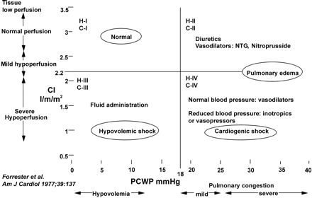
Figure 1 Clinical classification of the mode of heart failure (Forrester classification). H I-IV refers to haemodynamic severity, with reference figures for CI and pulmonary capillary pressures shown on the vertical and horizontal axes, respectively. C I-IV refers to clinical severity.
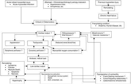
Figure 2 Pathophysiology of the syndrome of acute heart failure. Following acute critical events, LV deterioration occurs rapidly and requires urgent medical treatment. The pathophysiology of the syndrome of heart failure is summarized. Mechanical, haemodynamic and neurohormonal changes are similar but not identical to those observed in CHF. The time course of development or reversal of these changes varies considerably and strongly depends on the underlying cause of LV deterioration as well as pre-existing cardiovascular disease. However, changes develop rapidly and therefore AHF is considerably different to the syndrome of CHF.
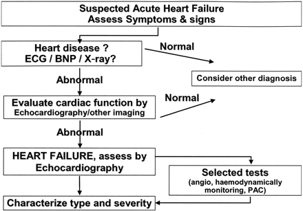
Figure 3 Diagnosis of AHF.
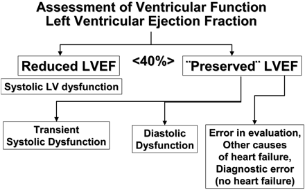
Figure 4 Assessment of LV function in AHF.
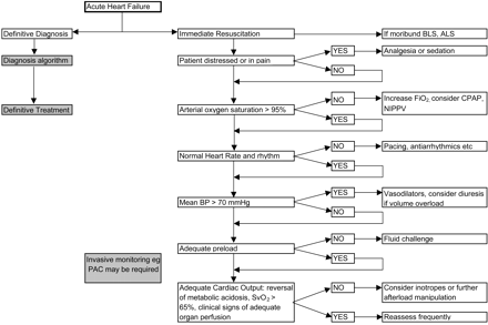
Figure 5 Immediate goals in treatment of the patients with AHF. In coronary patients mean blood pressure (mBP) should be higher to ensure coronary perfusion, mBP >70, or systolic >90 mmHg.
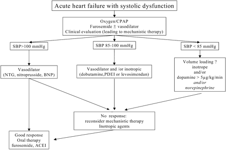
Figure 6 Rationale for inotropic drugs in AHF.
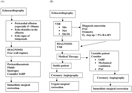
Figure 7 Algorithm: AHF in AMI. IABP=intra-aortic balloon pump; VSR=ventricular septal rupture; PAC=pulmonary artery catheterization; TEE=trans-esophageal echocardiography; EF=ejection fraction; MR=mitral regurgitation; IVS=intraventricular septum; SAM=systolic anterior movement; RA=right atrium; RV=right ventricle; PCI=percutaneous coronary intervention; Qp:Qs=pulmonary circulation volume:systemic circulation volume.
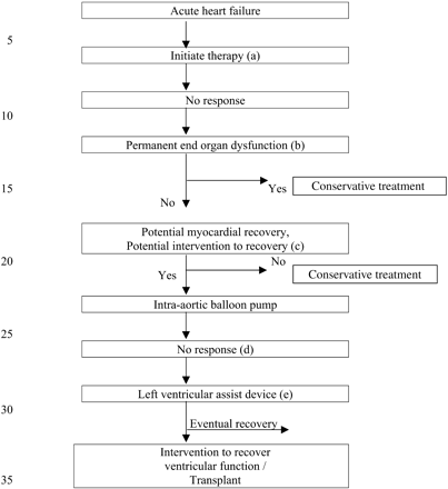
Figure 8 Selection of candidates for LV assist devices. (a): No response to conventional treatment of AHF, including appropriate use of diuretics and fluids, intravenous inotropics and vasodilators. (b): End organ dysfunction, including severe systemic disease, severe renal failure pulmonary disease or hepatic dysfunction, permanent central nervous injury. (c): Potential recovery of myocardial function or cardiac function, e.g. acute myocardial ischaemia, post-cardiotomy shock, acute myocarditis, acute valvular heart disease, or candidate to heart transplant. (d): Absence of clinical improvement after intra-aortic balloon pumping and mechanical ventilation. (e): Final indication may depend upon availability of device and experience of cardiovascular team.
Causes and precipitating factors in AHF
|
|
Causes and precipitating factors in AHF
|
|
Terminology and common clinical and haemodynamic characteristics
| Clinical status | Heart rate | SBP mmHg | CI L/min/m2 | PCWP mmHg | Congestion Killip/Forrester | Diuresis | Hypoperfusion | End organ hypoperfusion |
|---|---|---|---|---|---|---|---|---|
| I Acute decompensated congestive heart failure | +/− | Low normal/High | Low normal/High | Mild elevation | K II/F II | + | +/− | − |
| II Acute heart failure with hypertension/hypertensive crisis | Usually increased | High | +/− | >18 | K II-IV/FII-III | +/− | +/− | +, with CNS symptoms |
| III Acute heart failure with pulmonary oedema | + | Low normal | Low | Elevated | KIII/FII | + | +/− | − |
| IVa Cardiogenic shock*/low output syndrome | + | Low normal | Low, <2.2 | >16 | K III-IV/F I-III | low | + | + |
| IVb Severe cardiogenic shock | >90 | <90 | <1.8 | >18 | K IV/F IV | Very low | ++ | + |
| V High output failure | + | +/− | + | +/− | KII/FI-II | + | − | − |
| VI Right sided acute heart failure | Usually low | Low | Low | Low | F I | +/− | +/−, acute onset | +/− |
There are exceptions; the above values in table II are general rules.
*The differentation from low cardiac output syndrome is subjective and the clinical presentation may overlap these classifications.
SBP=systolic blood pressure; CI=cardiac index; PCWP=pulmonary capillary wedge pressure; CNS=central nervous system.
Laboratory tests in patients hospitalized with AHF
| Blood count | Always |
| Platelet count | Always |
| INR | If patient anticoagulated or in severe heart failure |
| CRP | Always |
| D-dimer | Always (may be falsely positive if CRP elevated or patient has been hospitalized for prolonged period) |
| Urea and Electrolytes (Na+, K+, Urea, Creatinine) | Always |
| Blood glucose | Always |
| CKMB, cardiac TnI/TnT | Always |
| Arterial blood gases | In severe heart failure, or in diabetic patients |
| Transaminases | To be considered |
| Urinanalysis | To be considered |
| Plasma BNP or NTproBNP | To be considered |
Other specific laboratory tests should be taken for differential diagnostic purposes or in order to identify end-organ dysfunction.
INR=international normalized ratio of thromboplastin time; TnI=troponin I; TnT=troponin T.
Goals of treatment of the patient with AHF
| Clinical |
| ↓ symptoms (dyspnoea and/or fatigue) |
| ↓ clinical signs |
| ↓ body weight |
| ↑ diuresis |
| ↑ oxygenation |
| Laboratory |
| Serum electrolyte normalization |
| ↓ BUN and/or creatinine |
| ↓ S-bilirubin |
| ↓ plasma BNP |
| Blood glucose normalization |
| Haemodynamic |
| ↓ pulmonary capillary wedge pressure to <18 mmHg |
| ↑ cardiac output and/or stroke volume |
| Outcome |
| ↓ length of stay in the intensive care unit |
| ↓ duration of hospitalization |
| ↑ time to hospital re-admission |
| ↓ mortality |
| Tolerability |
| Low rate of withdrawal from therapeutic measures |
| Low incidence of adverse effects |
BUN=blood urea nitrogen.
General therapeutic approach in AHF by findings on invasive haemodynamic monitoring
| Haemodynamic characteristic | Suggested therapeutic approach | ||||
|---|---|---|---|---|---|
| CI | Decreased | Decreased | Decreased | Decreased | Maintained |
| PCWP | Low | High or Normal | High | High | High |
| SBP mmHg | >85 | <85 | >85 | ||
| Outline of therapy | Fluid loading | Vasodilator (nitroprusside, NTG) fluid loading may become necessary | Consider inotropic agents (dobutamine, dopamine) and i.v. diuretics | Vasodilators (nitroprusside, NTG) and i.v. diuretics and consider inotrope (dobutamine, levosimendan, PDEI) | i.v. diuretics If SBP is low, vasoconstrictive inotropes |
In AHF patients: Decreased CI: <2.2 L/min/m2; PCWP: low if <14 mmHg, high if >18–20 mmHg.
Indications and dosing of vasodilators in AHF
| Vasodilator | Indication | Dosing | Main side effects | Other |
|---|---|---|---|---|
| Glyceryl trinitrate, 5-mononitrate | Acute heart failure, when blood pressure is adequate | Start 20 µg/min, increase to 200 µg/min | Hypotension, headache | Tolerance on continuous use |
| Isosorbide dinitrate | Acute heart failure, when blood pressure is adequate | Start with 1 mg/h, increase to 10 mg/h | Hypotension, headache | Tolerance on continuous use |
| Nitroprusside | Hypertensive crisis, cardiogenic shock combined with intoropes | 0.3–5µg/kg/min | Hypotension, isocyanate toxicity | Drug is light sensitive |
| Nesiritidea | Acute decompensated heart failure | Bolus 2 µg/kg + infusion 0.015–0.03 µg/kg/min | Hypotension |
aLimited sales approval in ESC countries.
Practical use of diuretics in AHF
| Start with individualized dose depending on clinical condition (see Table 8) |
| Titrate according to clinical response |
| Reduce dose when fluid retention is controlled |
| Monitor serum K+, Na+ and renal function at frequent intervals (every 1–2 days), according to diuretic response |
| Replace K+ and Mg+ loss |
| In case of diuretic resistance follow suggestions in Table 10 |
Diuretic dosing and administration
| Severity of fluid retention | Diuretic | Dose (mg) | Comments |
|---|---|---|---|
| Moderate | Furosemide, or | 20–40 | Oral or intravenous according to clinical symptoms |
| Bumetanide, or | 0.5–1.0 | Titrate dose according to clinical response | |
| Torasemide | 10–20 | Monitor Na+, K+, creatinine and blood pressure | |
| Severe | Furosemide, or Furosemide infusion | 40–100 | Intravenously |
| 5–40 mg/h | Better than very high bolus doses | ||
| Bumetanide, or Torasemide | 1–4 | Orally or intravenously | |
| 20–100 | Orally | ||
| Refractory to loop diuretics | Add HCTZ, or | 25–50 twice daily | Combination with loop diuretic better than very high dose of loop diuretics alone |
| Metolazone, or | 2.5–10 once daily | Metolazone more potent if creatinine clearance <30 mL/min | |
| Spironolactone | 25–50 once daily | Spironolactone best choice if patient not in renal failure and normal or low serum K+ | |
| In case of alkalosis | Acetazolamide | 0.5 | Intravenously |
| Refractory to loop diuretic and thiazides | Add dopamine for renal vasodilatation, or dobutamine as an inotropic agent | Consider ultrafiltration or haemodialysis if co-existing renal failure |
HCTZ=hydrochlorothiazide.
Causes of diuretic resistance
| Intravascular volume depletion |
| Neurohormonal activation |
| Rebound Na+ uptake after volume loss |
| Hypertrophy of distal nephron |
| Reduced tubular secretion (renal failure, NSAIDs) |
| Decreased renal perfusion (low output) |
| Impaired gut absorption of an oral diuretic |
| Non-compliance with drugs or diet (high sodium intake) |
Managing resistance to diuretics
| Restrict Na+/H2O intake and follow electrolytes113 |
| Volume repletion in cases of hypovolaemia113 |
| Increase dose and/or frequency of administration of diuretic109,116 |
| Use intravenous administration (more effective than oral)113 as bolus, or as intravenous infusion (more effective than high dose intravenous bolus)103–107,116 |
| Combine diuretic therapy108 |
| furosemide+HCTZ109 |
| furosemide+spironolactone111 |
| metolazone+furosemide (this combination is also active in renal failure)110,107 |
| Combine diuretic therapy with dopamine112, or dobutamine117 |
| Reduce the dose of ACE-inhibitor118 or use very low doses of ACE-inhibitor118,119 |
| Consider ultrafiltration or dialysis if response to above strategies ineffective120 |
Administration of positive inotropic agents
| Bolus | Infusion rate | |
|---|---|---|
| Dobutamine | No | 2 to 20 µg/kg/min (β+) |
| Dopamine | No | <3 µg/kg/min: renal effect (δ+) 3–5 µg/kg/min: inotropic (β+) >5 µg/kg/min: (β+), vasopressor (α+) |
| Milrinone | 25–75 µg/kg over 10–20 min | 0.375–0.75 µg/kg/min |
| Enoximone | 0.25–0.75 mg/kg | 1.25–7.5 µg/kg/min |
| Levosimendan | 12–24 µg/kga over 10 min | 0.1 µg/kg/min which can be decreased to 0.05 or increased to 0.2 µg/kg/min |
| Norepinephrine | No bolus | 0.2–1.0 µg/kg/min |
| Epinephrine | Bolus: 1 mg can be given i.v. at resuscitation, may be repeated after 3–5 min, endotracheal route is not favoured | 0.05–0.5 µg/kg/min |
aCurrent recommended dosing. In patients with hypotension, therapy should be started without a bolus.
Treatment of arrhythmias in acute heart failure
| Ventricular fibrillation or pulseless ventricular tachycardia | Defibrillate with 200–300–360J (preferably by biphasic defibrillation with a maximum of 200 J). If refractory to initial shocks inject epinephrine 1 mg or vasopressin 40 IU and/or amiodarone 150–300 mg as injection |
| Ventricular tachycardia | If patient is unstable cardiovert, if stable amiodarone or lidocaine can be given to achieve medical cardioversion. |
| Sinus tachycardia or supraventricular tachycardia | Use β-blocking agents when clinically and haemodynamically tolerated: Metoprolol 5 mg intravenously as a slow bolus (can be repeated if tolerated) Adenosine may be used to slow AV conduction or to cardiovert re-entrant tachycardia On rare occasions: Esmolol 0.5–1.0 mg/kg over 1 min, followed by infusion of 50–300 µg/kg/min, or Labetalol 1–2 mg bolus, followed by infusion of 1–2 mg/min (to total of 50–200 mg). Labetalol also indicated in AHF related to hypertensive crisis or phaeochoromcytoma, with 10-mg boluses, to a total dose of 300 mg. |
| Atrial fibrillation or flutter | Cardiovert if possible. Digoxin 0.125–0.25 mg iv, or β-blocking agent, or amiodarone, may be used to slow AV conduction. Amiodarone may induce medical cardioversion without compromising left ventricular haemodynamics. Patient should be heparinized. |
| Bradycardia | Atropine 0.25–0.5 mg iv, to total of 1–2 mg. As interim measure, isoproterenol 1 mg in 100 mL NaCl infused to a maximum of 75 mL/h (2–12 µg/min). If bradycardia is atropine-resistant, transcutaneous or transvenous pacing should be used as an interim measure. Theophylline may be used in AMI patients with atropine-resistant bradycardia with bolus of 0.25–0.5 mg/kg followed by infusion at 0.2–0.4 mg/kg/h |
Cardiac disorders and AHF requiring surgical treatment
| Cardiogenic shock after AMI in patients with multi-vessel ischaemic heart disease |
| Post-infarction ventricular septal defect |
| Free wall rupture |
| Acute decompensation of pre-existing heart valve disease |
| Prosthetic valve failure or thrombosis |
| Aortic aneurysm or aortic dissection rupture into the pericardial sac |
Acute mitral regurgitation from:
|
Acute aortic regurgitation from:
|
| Ruptured aneurysm of the sinus of Valsalva |
| Acute decompensation of chronic cardiomyopathy requiring support by mechanical assist devices |
Mechanical assist devices
| Device | System | Main indication | Comments |
|---|---|---|---|
| Extracorporeal | |||
| Continuous flow pumps | Several | Short term support | Easiest to use. Cheapest |
| Centrifugal pumps | Several | Extensive experience | |
| May include extracorporeal membrane oxygenators | |||
| Need for continuous monitoring | |||
| Patient bedridden | |||
| Pulsatile | Thoratec | Short term support | Easy to use. Not expensive |
| Abiomed | Post cardiotomy | Need for continuous monitoring | |
| LV and RV dysfunction | Patient bedridden | ||
| Bridge to heart transplant | |||
| Intracorporeal | |||
| Implantable, pulsatiles | Heart Mate | Long term use possible | Expensive |
| Novacor | Bridge to transplant | Patient may move and rehabilitate | |
| Bridge to recovery | |||
| Total artificial heart | No recovery expected | Experimental | |
| No transplant candidate | Limited experience | ||
| Alternative to transplant |
Remme WJ, Swedberg K. Guidelines for the diagnosis and treatment of chronic heart failure.
Guidelines on the diagnosis and treatment of acute heart failure. Available at http://www.escardio.org.
Effect of prophylactic amiodarone on mortality after acute myocardial infarction and in congestive heart failure: meta-analysis of individual data from 6500 patients in randomised trials. Amiodarone Trials Meta-Analysis Investigators.
McCullough PA, Philbin EF, Spertus JA et al. Confirmation of a heart failure epidemic: findings from the Resource Utilization Among Congestive Heart Failure (REACH) study.
Cleland JG, Swedberg K, Follath F et al. The EuroHeart Failure survey programme-a survey on the quality of care among patients with heart failure in Europe. Part 1: Patient characteristics and diagnosis.
Fox KF, Cowie MR, Wood DA et al. Coronary artery disease as the cause of incident heart failure in the population.
Al-Khadra AS, Salem DN, Rand WM et al. Warfarin anticoagulation and survival: a cohort analysis from the studies of left ventricular dysfunction.
Berry C, Murdoch DR, McMurray JJ. Economics of chronic heart failure.
The treatment of heart failure. Task Force of the Working Group on Heart Failure of the European Society of Cardiology.
Adams KF, Jr., Zannad F. Clinical definition and epidemiology of advanced heart failure.
Stevenson R, Ranjadayalan K, Wilkinson P et al. Short and long term prognosis of acute myocardial infarction since introduction of thrombolysis.
Roguin A, Behar D, Ben Ami H et al. Long-term prognosis of acute pulmonary oedema—an ominous outcome.
Krumholz MH P, EM, Tu N et al. The treatment target in acute decompensated heart failure.
Krumholz HM, Chen J, Murillo JE et al. Admission to hospitals with on-site cardiac catheterization facilities: impact on long-term costs and outcomes.
Cowie MR, Mosterd A, Wood DA et al. The epidemiology of heart failure.
McAlister FA, Lawson FM, Teo KK et al. A systematic review of randomized trials of disease management programs in heart failure.
Krumholz HM, Vaccarino V, Ellerbeck EF et al. Determinants of appropriate use of angiotensin-converting enzyme inhibitors after acute myocardial infarction in persons > or = 65 years of age.
Rich MW, Beckham V, Wittenberg C et al. A multidisciplinary intervention to prevent the readmission of elderly patients with congestive heart failure.
Nohria A TS, Fang JC, Lewis EF et al. Clinical assessment identifies hemodynamic profiles that predict outcomes in patients admitted with heart failure.
Grady KL, Dracub K, Kennedy G, Moser DK, Piano M, Stevensson LW. AHA Scientific Statement: Team management of patients with heart failure: a statement of health care professional from the cardiovascular nursing council of the American Heart Association.
Nohria A, Lewis E, Stevenson LW. Medical management of advanced heart failure.
Killip T, 3rd, Kimball JT. Treatment of myocardial infarction in a coronary care unit. A two year experience with 250 patients.
Forrester JS, Diamond GA, Swan HJ. Correlative classification of clinical and hemodynamic function after acute myocardial infarction.
Arnold JM, Braunwald E, Sandor T et al. Inotropic stimulation of reperfused myocardium with dopamine: effects on infarct size and myocardial function.
Marban E. Myocardial stunning and hibernation. The physiology behind the colloquialisms.
Bertrand ME, Simoons ML, Fox KA et al. Management of acute coronary syndromes in patients presenting without persistent ST-segment elevation.
Van de Werf F, Ardissino D, Betriu A et al. Management of acute myocardial infarction in patients presenting with ST-segment elevation. The Task Force on the Management of Acute Myocardial Infarction of the European Society of Cardiology.
Fuster V, Ryden LE, Asinger RW et al. ACC/AHA/ESC guidelines for the management of patients with atrial fibrillation. A report of the American College of Cardiology/American Heart Association Task Force on Practice Guidelines and the European Society of Cardiology Committee for Practice Guidelines and Policy Conferences (Committee to develop guidelines for the management of patients with atrial fibrillation) developed in collaboration with the North American Society of Pacing and Electrophysiology.
Maisel AS, Krishnaswamy P, Nowak RM et al. Rapid measurement of B-type natriuretic peptide in the emergency diagnosis of heart failure.
Dao Q, Krishnaswamy P, Kazanegra R et al. Utility of B-type natriuretic peptide in the diagnosis of congestive heart failure in an urgent-care setting.
Cowie MR, Jourdain P, Maisel A et al. Clinical applications of B-type natriuretic peptide (BNP) testing.
Capomolla S, Pozzoli M, Opasich C et al. Dobutamine and nitroprusside infusion in patients with severe congestive heart failure: hemodynamic improvement by discordant effects on mitral regurgitation, left atrial function, and ventricular function.
Tousignant CP, Walsh F, Mazer CD. The use of transesophageal echocardiography for preload assessment in critically ill patients.
Nagueh SF, Kopelen HA, Zoghbi WA. Feasibility and accuracy of Doppler echocardiographic estimation of pulmonary artery occlusive pressure in the intensive care unit.
Nishimura RA, Tajik AJ. Determination of left-sided pressure gradients by utilizing Doppler aortic and mitral regurgitant signals: validation by simultaneous dual catheter and Doppler studies.
Braunwald E, Antman EM, Beasley JW et al. ACC/AHA 2002 guideline update for the management of patients with unstable angina and non-ST-segment elevation myocardial infarction—summary article: a report of the American College of Cardiology/American Heart Association Task Force on Practice Guidelines (Committee on the Management of Patients With Unstable Angina).
Colucci WS, Elkayam U, Horton DP et al. Intravenous nesiritide, a natriuretic peptide, in the treatment of decompensated congestive heart failure. Nesiritide Study Group.
Torre-Amione G, Young JB, Durand J et al. Hemodynamic effects of tezosentan, an intravenous dual endothelin receptor antagonist, in patients with class III to IV congestive heart failure.
Follath F, Cleland JG, Just H et al. Efficacy and safety of intravenous levosimendan compared with dobutamine in severe low-output heart failure (the LIDO study): a randomised double-blind trial.
Steimle AE, Stevenson LW, Chelimsky-Fallick C et al. Sustained hemodynamic efficacy of therapy tailored to reduce filling pressures in survivors with advanced heart failure.
Gottlieb SS, Brater DC, Thomas I et al. BG9719 (CVT-124), an A1 adenosine receptor antagonist, protects against the decline in renal function observed with diuretic therapy.
Shah MR, Stinnett SS, McNulty SE et al. Hemodynamics as surrogate end points for survival in advanced heart failure: an analysis from FIRST.
Boyd O, Grounds RM, Bennett ED. A randomized clinical trial of the effect of deliberate perioperative increase of oxygen delivery on mortality in high-risk surgical patients.
Sandham JD, Hull RD, Brant RF et al. A randomized, controlled trial of the use of pulmonary-artery catheters in high-risk surgical patients.
Rhodes A, Cusack RJ, Newman PJ et al. A randomised, controlled trial of the pulmonary artery catheter in critically ill patients.
Mueller HS, Chatterjee K, Davis KB et al. ACC expert consensus document. Present use of bedside right heart catheterization in patients with cardiac disease.
van den Berghe G, Wouters P, Weekers F et al. Intensive insulin therapy in the critically ill patients.
Rawles JM, Kenmure AC. Controlled trial of oxygen in uncomplicated myocardial infarction.
Packer PM. Proposal for a new clinical end point to evaluate the efficacy of drugs and devices in the treatment of chronic heart failure.
Udelson JE, Smith WB, Hendrix GH et al. Acute hemodynamic effects of conivaptan, a dual V(1A) and V(2) vasopressin receptor antagonist, in patients with advanced heart failure.
Cuffe MS, Califf RM, Adams KF Jr., et al. Short-term intravenous milrinone for acute exacerbation of chronic heart failure: a randomized controlled trial.
Silver MA, Horton DP, Ghali JK et al. Effect of nesiritide versus dobutamine on short-term outcomes in the treatment of patients with acutely decompensated heart failure.
Fonarow GC, Stevenson LW, Walden JA et al. Impact of a comprehensive heart failure management program on hospital readmission and functional status of patients with advanced heart failure.
Marik PE. Pulmonary artery catheterization and esophageal doppler monitoring in the ICU.
Jardin F, Valtier B, Beauchet A et al. Invasive monitoring combined with two-dimensional echocardiographic study in septic shock.
Gore JM, Goldberg RJ, Spodick DH et al. A community-wide assessment of the use of pulmonary artery catheters in patients with acute myocardial infarction.
Zion MM, Balkin J, Rosenmann D et al. Use of pulmonary artery catheters in patients with acute myocardial infarction. Analysis of experience in 5,841 patients in the SPRINT Registry. SPRINT Study Group.
Connors AF Jr., Speroff T, Dawson NV et al. The effectiveness of right heart catheterization in the initial care of critically ill patients. SUPPORT Investigators.
Ivanov R, Allen J, Calvin JE. The incidence of major morbidity in critically ill patients managed with pulmonary artery catheters: a meta-analysis.
Wilson J, Woods I, Fawcett J et al. Reducing the risk of major elective surgery: randomised controlled trial of preoperative optimisation of oxygen delivery.
Rasanen J, Heikkila J, Downs J et al. Continuous positive airway pressure by face mask in acute cardiogenic pulmonary edema.
Bersten AD, Holt AW, Vedig AE et al. Treatment of severe cardiogenic pulmonary edema with continuous positive airway pressure delivered by face mask.
Lin M, Yang YF, Chiang HT et al. Reappraisal of continuous positive airway pressure therapy in acute cardiogenic pulmonary edema. Short-term results and long-term follow-up.
Takeda S, Nejima J, Takano T et al. Effect of nasal continuous positive airway pressure on pulmonary edema complicating acute myocardial infarction.
Kelly CA, Newby DE, McDonagh TA et al. Randomised controlled trial of continuous positive airway pressure and standard oxygen therapy in acute pulmonary oedema; effects on plasma brain natriuretic peptide concentrations.
Pang D, Keenan SP, Cook DJ et al. The effect of positive pressure airway support on mortality and the need for intubation in cardiogenic pulmonary edema: a systematic review.
Masip J, Betbese AJ, Paez J et al. Non-invasive pressure support ventilation versus conventional oxygen therapy in acute cardiogenic pulmonary oedema: a randomised trial.
Sharon A, Shpirer I, Kaluski E et al. High-dose intravenous isosorbide-dinitrate is safer and better than Bi-PAP ventilation combined with conventional treatment for severe pulmonary edema.
Mehta S, Jay GD, Woolard RH et al. Randomized, prospective trial of bilevel versus continuous positive airway pressure in acute pulmonary edema.
Lee G, DeMaria AN, Amsterdam EA et al. Comparative effects of morphine, meperidine and pentazocine on cardiocirculatory dynamics in patients with acute myocardial infarction.
Samama MM, Cohen AT, Darmon JY et al. A comparison of enoxaparin with placebo for the prevention of venous thromboembolism in acutely ill medical patients. Prophylaxis in Medical Patients with Enoxaparin Study Group.
Cotter G, Metzkor E, Kaluski E et al. Randomised trial of high-dose isosorbide dinitrate plus low-dose furosemide versus high-dose furosemide plus low-dose isosorbide dinitrate in severe pulmonary oedema.
Cohn JN, Franciosa JA. Vasodilator therapy of cardiac failure (second of two parts).
Jain P, Massie BM, Gattis WA et al. Current medical treatment for the exacerbation of chronic heart failure resulting in hospitalization.
Reves JG, Erdmann W, Mardis M et al. Evidence for existence of intramyocardial steal.
Colucci WS. Nesiritide for the treatment of decompensated heart failure.
Swedberg K, Held P, Kjekshus J et al. Effects of early administration of enalapril on mortality in patients with acute myocardial infarction. Results of the Cooperative New Scandinavian Enalapril Survival Study II (CONSENSUS II).
Ambrosioni E, Borghi C, Magnani B. The effect of the angiotensin-converting-enzyme inhibitor zofenopril on mortality and morbidity after anterior myocardial infarction. The Survival of Myocardial Infarction Long-Term Evaluation (SMILE) Study Investigators.
Latini R, Tognoni G, Maggioni AP et al. Clinical effects of early angiotensin-converting enzyme inhibitor treatment for acute myocardial infarction are similar in the presence and absence of aspirin: systematic overview of individual data from 96,712 randomized patients. Angiotensin-converting Enzyme Inhibitor Myocardial Infarction Collaborative Group.
Follath F. Do diuretics differ in terms of clinical outcome in congestive heart failure?
Wilson JR, Reichek N, Dunkman WB et al. Effect of diuresis on the performance of the failing left ventricle in man.
Johnson W, Omland T, Hall C et al. Neurohormonal activation rapidly decreases after intravenous therapy with diuretics and vasodilators for class IV heart failure.
Brater DC. Resistance to loop diuretics. Why it happens and what to do about it.
Cotter G, Metzkor E, Kaluski E et al. Randomised trial of high-dose isosorbide dinitrate plus low-dose furosemide versus high-dose furosemide plus low-dose isosorbide dinitrate in severe pulmonary oedema [see comments].
Gardtman M, Waagstein L, Karlsson T et al. Has an intensified treatment in the ambulance of patients with acute severe left heart failure improved the outcome?
Sacchetti A, Ramoska E, Moakes ME et al. Effect of ED management on ICU use in acute pulmonary edema.
Kramer WG, Smith WB, Ferguson J et al. Pharmacodynamics of torsemide administered as an intravenous injection and as a continuous infusion to patients with congestive heart failure.
Lahav M, Regev A, Ra'anani P et al. Intermittent administration of furosemide vs continuous infusion preceded by a loading dose for congestive heart failure.
Pivac N, Rumboldt Z, Sardelic S et al. Diuretic effects of furosemide infusion versus bolus injection in congestive heart failure.
van Meyel JJ, Smits P, Dormans T et al. Continuous infusion of furosemide in the treatment of patients with congestive heart failure and diuretic resistance.
Channer KS, McLean KA, Lawson-Matthew P et al. Combination diuretic treatment in severe heart failure: a randomised controlled trial.
Dormans TP, Gerlag PG, Russel FG et al. Combination diuretic therapy in severe congestive heart failure.
Ellison DH. Diuretic therapy and resistance in congestive heart failure.
Kiyingi A, Field MJ, Pawsey CC et al. Metolazone in treatment of severe refractory congestive cardiac failure.
van Vliet AA, Donker AJ, Nauta JJ et al. Spironolactone in congestive heart failure refractory to high-dose loop diuretic and low-dose angiotensin-converting enzyme inhibitor.
Cotter G, Weissgarten J, Metzkor E et al. Increased toxicity of high-dose furosemide versus low-dose dopamine in the treatment of refractory congestive heart failure.
Kramer BK, Schweda F, Riegger GA. Diuretic treatment and diuretic resistance in heart failure.
Neuberg GW, Miller AB, O'Connor CM et al. Diuretic resistance predicts mortality in patients with advanced heart failure.
Wakelkamp M, Alvan G, Gabrielsson J et al. Pharmacodynamic modeling of furosemide tolerance after multiple intravenous administration.
Dormans TP, van Meyel JJ, Gerlag PG et al. Diuretic efficacy of high dose furosemide in severe heart failure: bolus injection versus continuous infusion.
Maxwell AP, Ong HY, Nicholls DP. Influence of progressive renal dysfunction in chronic heart failure.
Marik PE, Kussman BD, Lipman J et al. Acetazolamide in the treatment of metabolic alkalosis in critically ill patients.
Sharpe N. Beta-blockers in heart failure. Future directions.
Furberg CD. Overview of completed sudden death trials: US experience.
Yusuf S, Peto R, Lewis J et al. Beta blockade during and after myocardial infarction: an overview of the randomized trials.
Herlitz J, Waagstein F, Lindqvist J et al. Effect of metoprolol on the prognosis for patients with suspected acute myocardial infarction and indirect signs of congestive heart failure (a subgroup analysis of the Goteborg Metoprolol Trial).
Herlitz J, Elmfeldt D, Hjalmarson A et al. Effect of metoprolol on indirect signs of the size and severity of acute myocardial infarction.
Witchitz S, Cohen-Solal A, Dartois N et al. Treatment of heart failure with celiprolol, a cardioselective beta blocker with beta-2 agonist vasodilatory properties. The CELICARD Group.
Held PH, Corbeij HM, Dunselman P et al. Hemodynamic effects of metoprolol in acute myocardial infarction. A randomized, placebo-controlled multicenter study.
Katz AM. Potential deleterious effects of inotropic agents in the therapy of chronic heart failure.
Packer M. The development of positive inotropic agents for chronic heart failure: how have we gone astray?
O'Connor CM, Gattis WA, Uretsky BF et al. Continuous intravenous dobutamine is associated with an increased risk of death in patients with advanced heart failure: Insights from the Flolan International Randomized Survival Trial (FIRST).
Thackray S, Easthaugh J, Freemantle N et al. The effectiveness and relative effectiveness of intravenous inotropic drugs acting through the adrenergic pathway in patients with heart failure—a meta-regression analysis.
Goldberg LI, McDonald RH, Jr., Zimmerman AM. Sodium diuresis produced by dopamine in patients with congestive heart failure.
Colucci WS, Wright RF, Braunwald E. New positive inotropic agents in the treatment of congestive heart failure. Mechanisms of action and recent clinical developments. 1.
Maskin CS, Ocken S, Chadwick B et al. Comparative systemic and renal effects of dopamine and angiotensin-converting enzyme inhibition with enalapril in patients with heart failure.
Metra M, Missale C, Spano PF et al. Dopaminergic drugs in congestive heart failure: hemodynamic and neuroendocrine responses to ibopamine, dopamine, and dihydroergotoxine.
Leier CV, Binkley PF. Parenteral inotropic support for advanced congestive heart failure.
Fowler MB, Laser JA, Hopkins GL et al. Assessment of the beta-adrenergic receptor pathway in the intact failing human heart: progressive receptor down-regulation and subsensitivity to agonist response.
Feldman MD, Copelas L, Gwathmey JK et al. Deficient production of cyclic AMP: pharmacologic evidence of an important cause of contractile dysfunction in patients with end-stage heart failure.
Colucci WS, Denniss AR, Leatherman GF et al. Intracoronary infusion of dobutamine to patients with and without severe congestive heart failure. Dose-response relationships, correlation with circulating catecholamines, and effect of phosphodiesterase inhibition.
Colucci WS, Wright RF, Jaski BE et al. Milrinone and dobutamine in severe heart failure: differing hemodynamic effects and individual patient responsiveness.
Lowes BD, Tsvetkova T, Eichhorn EJ et al. Milrinone versus dobutamine in heart failure subjects treated chronically with carvedilol.
Metra M, Nodari S, D'Aloia A et al. Beta-blocker therapy influences the hemodynamic response to inotropic agents in patients with heart failure: a randomized comparison of dobutamine and enoximone before and after chronic treatment with metoprolol or carvedilol.
Gilbert EM, Hershberger RE, Wiechmann RJ et al. Pharmacologic and hemodynamic effects of combined beta-agonist stimulation and phosphodiesterase inhibition in the failing human heart.
Levine TB, Levine AB, Elliott WG et al. Dobutamine as bridge to angiotensin-converting enzyme inhibitor-nitrate therapy in endstage heart failure.
Caldicott LD, Hawley K, Heppell R et al. Intravenous enoximone or dobutamine for severe heart failure after acute myocardial infarction: a randomized double-blind trial.
Burger AJ, Horton DP, LeJemtel T et al. Effect of nesiritide (B-type natriuretic peptide) and dobutamine on ventricular arrhythmias in the treatment of patients with acutely decompensated congestive heart failure: the PRECEDENT study.
Schulz R, Rose J, Martin C et al. Development of short-term myocardial hibernation. Its limitation by the severity of ischemia and inotropic stimulation.
Colucci WS, Wright RF, Braunwald E. New positive inotropic agents in the treatment of congestive heart failure. Mechanisms of action and recent clinical developments. 2.
Bohm M, Deutsch HJ, Hartmann D et al. Improvement of postreceptor events by metoprolol treatment in patients with chronic heart failure.
Loh E, Elkayam U, Cody R et al. A randomized multicenter study comparing the efficacy and safety of intravenous milrinone and intravenous nitroglycerin in patients with advanced heart failure.
Kivikko M, Lehtonen L, Colucci WS. Sustained hemodynamic effects of intravenous levosimendan.
Innes CA, Wagstaff AJ. Levosimendan: A review of its use in the management of acute decompensated heart failure.
Nieminen MS, Lilleberg J, Leikola-Pelho T et al. Dose related responses of a new calcium-sensitizer, simendan, in man.
Nieminen MS, Akkila J, Hasenfuss G et al. Hemodynamic and neurohumoral effects of continuous infusion of levosimendan in patients with congestive heart failure.
Slawsky MT, Colucci WS, Gottlieb SS et al. Acute hemodynamic and clinical effects of levosimendan in patients with severe heart failure. Study Investigators.
Cleland JG, McGowan J. Levosimendan: a new era for inodilator therapy for heart failure?
Bohm M, Beuckelmann D, Brown L et al. Reduction of beta-adrenoceptor density and evaluation of positive inotropic responses in isolated, diseased human myocardium.
Bohm M, La Rosee K, Schmidt U et al. Force-frequency relationship and inotropic stimulation in the nonfailing and failing human myocardium: implications for the medical treatment of heart failure.
The effect of digoxin on mortality and morbidity in patients with heart failure. The Digitalis Investigation Group.
Hood WB Jr, Dans AL, Guyatt GH et al. Digitalis for treatment of congestive heart failure in patients in sinus rhythm. The Cochrane Database of Systematic Reviews 2004 Issue 4.
Ratshin RA, Rackley CE, Russell RO, Jr. Hemodynamic evaluation of left ventricular function in shock complicating myocardial infarction.
Lee DC, Johnson RA, Bingham JB et al. Heart failure in outpatients: a randomized trial of digoxin versus placebo.
Rahimtoola SH, Sinno MZ, Chuquimia R et al. Effects of ouabain on impaired left ventricular function in acute myocardial infarction.
Spargias KS, Hall AS, Ball SG. Safety concerns about digoxin after acute myocardial infarction.
Varonkov Y, Shell WE, Smirnov V et al. Augmentation of serum CPK activity by digitalis in patients with acute myocardial infarction.
McClement BM AA. Value of signal-averaged electrocardiolography, radionuclide ventriculopathy, Holter monitoring and clinical variables for prediction of arrhythmic events in survivors of acute myocardial infarction in the thrombolytic era.
Khand AU, Rankin AC, Kaye GC et al. Systematic review of the management of atrial fibrillation in patients with heart failure.
Wong SC, Sanborn T, Sleeper LA et al. Angiographic findings and clinical correlates in patients with cardiogenic shock complicating acute myocardial infarction: a report from the SHOCK Trial Registry. SHould we emergently revascularize Occluded Coronaries for cardiogenic shocK?
Malmberg K, Norhammar A, Wedel H et al. Glycometabolic state at admission: important risk marker of mortality in conventionally treated patients with diabetes mellitus and acute myocardial infarction: long-term results from the Diabetes and Insulin-Glucose Infusion in Acute Myocardial Infarction (DIGAMI) study.
Hochman JS, Buller CE, Sleeper LA et al. Cardiogenic shock complicating acute myocardial infarction—etiologies, management and outcome: a report from the SHOCK Trial Registry. Should we emergently revascularize occluded Coronaries for cardiogenic shock?
Menon V, Slater JN, White HD et al. Acute myocardial infarction complicated by systemic hypoperfusion without hypotension: report of the SHOCK trial registry.
Sexton DJ, Spelman D. Current best practices and guidelines. Assessment and management of complications in infective endocarditis.
Olaison L, Pettersson G. Current best practices and guidelines. Indications for surgical intervention in infective endocarditis.
Houpikian P, Raoult D. Diagnostic methods. Current best practices and guidelines for identification of difficult-to-culture pathogens in infective endocarditis.
Towns ML, Reller LB. Diagnostic methods. Current best practices and guidelines for isolation of bacteria and fungi in infective endocarditis.
Conti CR. Endocarditis prophylaxis yes: endocarditis prophylaxis no.
Lengyel M, Fuster V, Keltai M et al. Guidelines for management of left-sided prosthetic valve thrombosis: a role for thrombolytic therapy. Consensus Conference on Prosthetic Valve Thrombosis.
Bonow RO, Carabello B, de Leon AC et al. ACC/AHA Guidelines for the Management of Patients With Valvular Heart Disease. Executive Summary. A report of the American College of Cardiology/American Heart Association Task Force on Practice Guidelines (Committee on Management of Patients with Valvular Heart Disease).
Alpert JS. The thrombosed prosthetic valve: current recommendations based on evidence from the literature.
Ozkan M, Kaymaz C, Kirma C et al. Intravenous thrombolytic treatment of mechanical prosthetic valve thrombosis: a study using serial transesophageal echocardiography.
Hering D, Piper C, Horstkotte D. Management of prosthetic valve thrombosis.
Roudaut R, Lafitte S, Roudaut MF et al. Fibrinolysis of mechanical prosthetic valve thrombosis: a single-center study of 127 cases.
Erbel R, Alfonso F, Boileau C et al. Diagnosis and management of aortic dissection.
Angeja BG, Grossman W. Evaluation and management of diastolic heart failure.
Burkhoff D, Maurer MS, Packer M. Heart failure with a normal ejection fraction: is it really a disorder of diastolic function?
Dalrymple-Hay MJ, Monro JL, Livesey SA et al. Postinfarction ventricular septal rupture: the Wessex experience.
Crenshaw BS, Granger CB, Birnbaum Y et al. Risk factors, angiographic patterns, and outcomes in patients with ventricular septal defect complicating acute myocardial infarction. GUSTO-I (Global Utilization of Streptokinase and TPA for Occluded Coronary Arteries) Trial Investigators.
Boersma E, Poldermans D, Bax JJ et al. Predictors of cardiac events after major vascular surgery: Role of clinical characteristics, dobutamine echocardiography, and beta-blocker therapy.
Anand IS, Chugh SS. Mechanisms and management of renal dysfunction in heart failure.
Weinfeld MS, Chertow GM, Stevenson LW. Aggravated renal dysfunction during intensive therapy for advanced chronic heart failure.
Hillege HL, Girbes AR, de Kam PJ et al. Renal function, neurohormonal activation, and survival in patients with chronic heart failure.
Leier CV, Dei Cas L, Metra M. Clinical relevance and management of the major electrolyte abnormalities in congestive heart failure: hyponatremia, hypokalemia, and hypomagnesemia.
Agostoni PG, Marenzi GC, Sganzerla P et al. Lung-heart interaction as a substrate for the improvement in exercise capacity after body fluid volume depletion in moderate congestive heart failure.
Sharma A, Hermann DD, Mehta RL. Clinical benefit and approach of ultrafiltration in acute heart failure.
Tepel M, van der Giet M, Schwarzfeld C et al. Prevention of radiographic-contrast-agent-induced reductions in renal function by acetylcysteine.
Briguori C, Manganelli F, Scarpato P et al. Acetylcysteine and contrast agent-associated nephrotoxicity.
Chu VL, Cheng JW. Fenoldopam in the prevention of contrast media-induced acute renal failure.
Marenzi G, Marana I, Lauri G et al. The prevention of radiocontrast-agent-induced nephropathy by hemofiltration.
Kindman LA, Vagelos RH, Willson K et al. Abnormalities of pulmonary function in patients with congestive heart failure, and reversal with ipratropium bromide.
Kitchen JB, 3rd, Kastor JA. Pacing in acute myocardial infarction—indications, methods, hazards, and results.
Priori SG, Aliot E, Blomstrom-Lundqvist C et al. Task Force on Sudden Cardiac Death, European Society of Cardiology.
Monsieurs KG, Handley AJ, Bossaert LL. European Resuscitation Council Guidelines 2000 for Automated External Defibrillation. A statement from the Basic Life Support and Automated External Defibrillation Working Group(1) and approved by the Executive Committee of the European Resuscitation Council.
Guidelines 2000 for Cardiopulmonary Resuscitation and Emergency Cardiovascular Care. Part 3: adult basic life support. The American Heart Association in collaboration with the International Liaison Committee on Resuscitation.
Mathew J, Hunsberger S, Fleg J et al. Incidence, predictive factors, and prognostic significance of supraventricular tachyarrhythmias in congestive heart failure.
Effect of metoprolol CR/XL in chronic heart failure: Metoprolol CR/XL Randomised Intervention Trial in Congestive Heart Failure (MERIT-HF).
Pedersen OD, Bagger H, Keller N et al. Efficacy of dofetilide in the treatment of atrial fibrillation-flutter in patients with reduced left ventricular function: a Danish investigations of arrhythmia and mortality on dofetilide (diamond) substudy.
Blomstrom-Lundqvist C, Scheinman MM, Aliot EM et al. ACC/AHA/ESC guidelines for the management of patients with supraventricular arrhythmias—executive summary. A report of the American College of Cardiology/American Heart Association Task Force on Practice Guidelines and the European Society of Cardiology Committee for Practice Guidelines (Writing Committee to Develop Guidelines for the Management of Patients with Supraventricular Arrhythmias) developed in collaboration with NASPE-Heart Rhythm Society.
Hebbar AK, Hueston WJ. Management of common arrhythmias: Part I. Supraventricular arrhythmias.
The pre-hospital management of acute heart attacks. Recommendations of a Task Force of the The European Society of Cardiology and The European Resuscitation Council.
Ellison KE, Stevenson WG, Sweeney MO et al. Catheter ablation for hemodynamically unstable monomorphic ventricular tachycardia.
Pohjola-Sintonen S, Muller JE, Stone PH et al. Ventricular septal and free wall rupture complicating acute myocardial infarction: experience in the Multicenter Investigation of Limitation of Infarct Size.
London RE LS. The electrocardiographic signs of acute hemopericardium.
Lopez-Sendon J, Gonzalez A, Lopez de Sa E et al. Diagnosis of subacute ventricular wall rupture after acute myocardial infarction: sensitivity and specificity of clinical, hemodynamic and echocardiographic criteria.
Zamorano J, Moreno R, Almeria C et al. Left ventricular free wall rupture during dobutamine stress echocardiography.
Deja MA, Szostek J, Widenka K et al. Post infarction ventricular septal defect—can we do better?
Ryan TJ, Antman EM, Brooks NH et al. 1999 update: ACC/AHA Guidelines for the Management of Patients with Acute Myocardial Infarction: Executive Summary and Recommendations: A report of the American College of Cardiology/American Heart Association Task Force on Practice Guidelines (Committee on Management of Acute Myocardial Infarction).
Haley JH, Sinak LJ, Tajik AJ et al. Dynamic left ventricular outflow tract obstruction in acute coronary syndromes: an important cause of new systolic murmur and cardiogenic shock.
Thompson CR, Buller CE, Sleeper LA et al. Cardiogenic shock due to acute severe mitral regurgitation complicating acute myocardial infarction: a report from the SHOCK Trial Registry. Should we use emergently revascularize Occluded Coronaries in cardiogenic shocK?
Tavakoli R, Weber A, Brunner-La Rocca H et al. Results of surgery for irreversible moderate to severe mitral valve regurgitation secondary to myocardial infarction.
Waksman R, Weiss AT, Gotsman MS et al. Intra-aortic balloon counterpulsation improves survival in cardiogenic shock complicating acute myocardial infarction.
Stevenson LW, Kormos RL. Mechanical Cardiac Support 2000: Current applications and future trial design.
Goldstein DJ, Oz MC, Rose EA. Implantable left ventricular assist devices.
Delgado DH, Rao V, Ross HJ et al. Mechanical circulatory assistance: state of art.
Bartlett RH, Roloff DW, Custer JR et al. Extracorporeal life support: the University of Michigan experience.
Author notes
Members of the European Society of Intensive Care Medicine

