


Table of Contents
Abbreviations and acronyms
Definition of myocardial infarction
Criteria for acute myocardial infarction
Criteria for prior myocardial infarction
Introduction
Pathological characteristics of myocardial ischaemia and infarction
Biomarker detection of myocardial injury with necrosis
Clinical features of myocardial ischaemia and infarction
Clinical classification of myocardial infarction
Spontaneous myocardial infarction (MI type 1)
Myocardial infarction secondary to an ischaemic imbalance (MI type 2)
Cardiac death due to myocardial infarction (MI type 3)
Myocardial infarction associated with revascularization procedures (MI types 4 and 5)
Electrocardiographic detection of myocardial infarction
Prior myocardial infarction
Silent myocardial infarction
Conditions that confound the ECG diagnosis of myocardial infarction
Imaging techniques
Echocardiography
Radionuclide imaging
Magnetic resonance imaging
Computed tomography
Applying imaging in acute myocardial infarction
Applying imaging in late presentation of myocardial infarction
Diagnostic criteria for myocardial infarction with PCI (MI type 4)
Diagnostic criteria for myocardial infarction with CABG (MI type 5)
Assessment of MI in patients undergoing other cardiac procedures
Myocardial infarction associated with non-cardiac procedures
Myocardial infarction in the intensive care unit
Recurrent myocardial infarction
Reinfarction
Myocardial injury or infarction associated with heart failure
Application of MI in clinical trials and quality assurance programmes
Public policy implications of the adjustment of the MI definition
Global perspectives of the definition of myocardial infarction
Conflicts of interest
Acknowledgements
References
Abbreviations and acronyms
- ACCF
American College of Cardiology Foundation
- ACS
acute coronary syndrome
- AHA
American Heart Association
- CAD
coronary artery disease
- CABG
coronary artery bypass grafting
- CKMB
creatine kinase MB isoform
- cTn
cardiac troponin
- CT
computed tomography
- CV
coefficient of variation
- ECG
electrocardiogram
- ESC
European Society of Cardiology
- FDG
fluorodeoxyglucose
- h
hour(s)
- HF
heart failure
- LBBB
left bundle branch block
- LV
left ventricle
- LVH
left ventricular hypertrophy
- MI
myocardial infarction
- mIBG
meta-iodo-benzylguanidine
- min
minute(s)
- MONICA
Multinational MONItoring of trends and determinants in CArdiovascular disease)
- MPS
myocardial perfusion scintigraphy
- MRI
magnetic resonance imaging
- mV
millivolt(s)
- ng/L
nanogram(s) per litre
- Non-Q MI
non-Q wave myocardial infarction
- NSTEMI
non-ST-elevation myocardial infarction
- PCI
percutaneous coronary intervention
- PET
positron emission tomography
- pg/mL
pictogram(s) per millilitre
- Q wave MI
Q wave myocardial infarction
- RBBB
right bundle branch block
- sec
second(s)
- SPECT
single photon emission computed tomography
- STEMI
ST elevation myocardial infarction
- ST–T
ST-segment –T wave
- URL
upper reference limit
- WHF
World Heart Federation
- WHO
World Health Organization
Introduction
Myocardial infarction (MI) can be recognised by clinical features, including electrocardiographic (ECG) findings, elevated values of biochemical markers (biomarkers) of myocardial necrosis, and by imaging, or may be defined by pathology. It is a major cause of death and disability worldwide. MI may be the first manifestation of coronary artery disease (CAD) or it may occur, repeatedly, in patients with established disease. Information on MI rates can provide useful information regarding the burden of CAD within and across populations, especially if standardized data are collected in a manner that distinguishes between incident and recurrent events. From the epidemiological point of view, the incidence of MI in a population can be used as a proxy for the prevalence of CAD in that population. The term ‘myocardial infarction’ may have major psychological and legal implications for the individual and society. It is an indicator of one of the leading health problems in the world and it is an outcome measure in clinical trials, observational studies and quality assurance programmes. These studies and programmes require a precise and consistent definition of MI.
In the past, a general consensus existed for the clinical syndrome designated as MI. In studies of disease prevalence, the World Health Organization (WHO) defined MI from symptoms, ECG abnormalities and cardiac enzymes. However, the development of ever more sensitive and myocardial tissue-specific cardiac biomarkers and more sensitive imaging techniques now allows for detection of very small amounts of myocardial injury or necrosis. Additionally, the management of patients with MI has significantly improved, resulting in less myocardial injury and necrosis, in spite of a similar clinical presentation. Moreover, it appears necessary to distinguish the various conditions which may cause MI, such as ‘spontaneous’ and ‘procedure-related’ MI. Accordingly, physicians, other healthcare providers and patients require an up-to-date definition of MI.
In 2000, the First Global MI Task Force presented a new definition of MI, which implied that any necrosis in the setting of myocardial ischaemia should be labelled as MI.1 These principles were further refined by the Second Global MI Task Force, leading to the Universal Definition of Myocardial Infarction Consensus Document in 2007, which emphasized the different conditions which might lead to an MI.2 This document, endorsed by the European Society of Cardiology (ESC), the American College of Cardiology Foundation (ACCF), the American Heart Association (AHA), and the World Heart Federation (WHF), has been well accepted by the medical community and adopted by the WHO.3 However, the development of even more sensitive assays for markers of myocardial necrosis mandates further revision, particularly when such necrosis occurs in the setting of the critically ill, after percutaneous coronary procedures or after cardiac surgery. The Third Global MI Task Force has continued the Joint ESC/ACCF/AHA/WHF efforts by integrating these insights and new data into the current document, which now recognizes that very small amounts of myocardial injury or necrosis can be detected by biochemical markers and/or imaging.
Pathological characteristics of myocardial ischaemia and infarction
MI is defined in pathology as myocardial cell death due to prolonged ischaemia. After the onset of myocardial ischaemia, histological cell death is not immediate, but takes a finite period of time to develop—as little as 20 min, or less in some animal models.4 It takes several hours before myocardial necrosis can be identified by macroscopic or microscopic post-mortem examination. Complete necrosis of myocardial cells at risk requires at least 2–4 h, or longer, depending on the presence of collateral circulation to the ischaemic zone, persistent or intermittent coronary arterial occlusion, the sensitivity of the myocytes to ischaemia, pre-conditioning, and individual demand for oxygen and nutrients.2 The entire process leading to a healed infarction usually takes at least 5–6 weeks. Reperfusion may alter the macroscopic and microscopic appearance.
Biomarker detection of myocardial injury with necrosis
Myocardial injury is detected when blood levels of sensitive and specific biomarkers such as cTn or the MB fraction of creatine kinase (CKMB) are increased.2 Cardiac troponin I and T are components of the contractile apparatus of myocardial cells and are expressed almost exclusively in the heart. Although elevations of these biomarkers in the blood reflect injury leading to necrosis of myocardial cells, they do not indicate the underlying mechanism.5 Various possibilities have been suggested for release of structural proteins from the myocardium, including normal turnover of myocardial cells, apoptosis, cellular release of troponin degradation products, increased cellular wall permeability, formation and release of membranous blebs, and myocyte necrosis.6 Regardless of the pathobiology, myocardial necrosis due to myocardial ischaemia is designated as MI.
Also, histological evidence of myocardial injury with necrosis may be detectable in clinical conditions associated with predominantly non-ischaemic myocardial injury. Small amounts of myocardial injury with necrosis may be detected, which are associated with heart failure (HF), renal failure, myocarditis, arrhythmias, pulmonary embolism or otherwise uneventful percutaneous or surgical coronary procedures. These should not be labelled as MI or a complication of the procedures, but rather as myocardial injury, as illustrated in Figure 1. It is recognized that the complexity of clinical circumstances may sometimes render it difficult to determine where individual cases may lie within the ovals of Figure 1. In this setting, it is important to distinguish acute causes of cTn elevation, which require a rise and/or fall of cTn values, from chronic elevations that tend not to change acutely. A list of such clinical circumstances associated with elevated values of cTn is presented in Table 1. The multifactorial contributions resulting in the myocardial injury should be described in the patient record.
This illustration shows various clinical entities: for example, renal failure, heart failure, tachy- or bradyarrhythmia, cardiac or non-cardiac procedures that can be associated with myocardial injury with cell death marked by cardiac troponin elevation. However, these entities can also be associated with myocardial infarction in case of clinical evidence of acute myocardial ischaemia with rise and/or fall of cardiac troponin.
The preferred biomarker—overall and for each specific category of MI—is cTn (I or T), which has high myocardial tissue specificity as well as high clinical sensitivity. Detection of a rise and/or fall of the measurements is essential to the diagnosis of acute MI.7 An increased cTn concentration is defined as a value exceeding the 99th percentile of a normal reference population [upper reference limit (URL)]. This discriminatory 99th percentile is designated as the decision level for the diagnosis of MI and must be determined for each specific assay with appropriate quality control in each laboratory.8,9 The values for the 99th percentile URL defined by manufacturers, including those for many of the high-sensitivity assays in development, can be found in the package inserts for the assays or in recent publications.10,11,12
Values should be presented as nanograms per litre (ng/L) or picograms per millilitre (pg/mL) to make whole numbers. Criteria for the rise of cTn values are assay-dependent but can be defined from the precision profile of each individual assay, including high-sensitivity assays.10,11 Optimal precision, as described by coefficient of variation (CV) at the 99th percentile URL for each assay, should be defined as ≤10%. Better precision (CV ≤10%) allows for more sensitive assays and facilitates the detection of changing values.13 The use of assays that do not have optimal precision (CV >10% at the 99th percentile URL) makes determination of a significant change more difficult but does not cause false positive results. Assays with CV >20% at the 99th percentile URL should not be used.13 It is acknowledged that pre-analytic and analytic problems can induce elevated and reduced values of cTn.10,11
Blood samples for the measurement of cTn should be drawn on first assessment and repeated 3–6 h later. Later samples are required if further ischaemic episodes occur, or when the timing of the initial symptoms is unclear.14 To establish the diagnosis of MI, a rise and/or fall in values with at least one value above the decision level is required, coupled with a strong pre-test likelihood. The demonstration of a rising and/or falling pattern is needed to distinguish acute- from chronic elevations in cTn concentrations that are associated with structural heart disease.10,11,15–19 For example, patients with renal failure or HF can have significant chronic elevations in cTn. These elevations can be marked, as seen in many patients with MI, but do not change acutely.7 However, a rising or falling pattern is not absolutely necessary to make the diagnosis of MI if a patient with a high pre-test risk of MI presents late after symptom onset; for example, near the peak of the cTn time-concentration curve or on the slow-declining portion of that curve, when detecting a changing pattern can be problematic. Values may remain elevated for 2 weeks or more following the onset of myocyte necrosis.10
Sex-dependent values may be recommended for high-sensitivity troponin assays.20,21 An elevated cTn value (>99th percentile URL), with or without a dynamic pattern of values or in the absence of clinical evidence of ischaemia, should prompt a search for other diagnoses associated with myocardial injury, such as myocarditis, aortic dissection, pulmonary embolism, or HF. Renal failure and other more non-ischaemic chronic disease states, that can be associated with elevated cTn levels, are listed in Table 1.10,11
If a cTn assay is not available, the best alternative is CKMB (measured by mass assay). As with troponin, an increased CKMB value is defined as a measurement above the 99th percentile URL, which is designated as the decision level for the diagnosis of MI.22 Sex-specific values should be employed.22
Clinical features of myocardial ischaemia and infarction
Onset of myocardial ischaemia is the initial step in the development of MI and results from an imbalance between oxygen supply and demand. Myocardial ischaemia in a clinical setting can usually be identified from the patient's history and from the ECG. Possible ischaemic symptoms include various combinations of chest, upper extremity, mandibular or epigastric discomfort (with exertion or at rest) or an ischaemic equivalent such as dyspnoea or fatigue. The discomfort associated with acute MI usually lasts >20 min. Often, the discomfort is diffuse—not localized, nor positional, nor affected by movement of the region—and it may be accompanied by diaphoresis, nausea or syncope. However, these symptoms are not specific for myocardial ischaemia. Accordingly, they may be misdiagnosed and attributed to gastrointestinal, neurological, pulmonary or musculoskeletal disorders. MI may occur with atypical symptoms—such as palpitations or cardiac arrest—or even without symptoms; for example in women, the elderly, diabetics, or post-operative and critically ill patients.2 Careful evaluation of these patients is advised, especially when there is a rising and/or falling pattern of cardiac biomarkers.
Clinical classification of myocardial infarction
For the sake of immediate treatment strategies, such as reperfusion therapy, it is usual practice to designate MI in patients with chest discomfort, or other ischaemic symptoms that develop ST elevation in two contiguous leads (see ECG section), as an ‘ST elevation MI’ (STEMI). In contrast, patients without ST elevation at presentation are usually designated as having a ‘non-ST elevation MI’ (NSTEMI). Many patients with MI develop Q waves (Q wave MI), but others do not (non-Q MI). Patients without elevated biomarker values can be diagnosed as having unstable angina. In addition to these categories, MI is classified into various types, based on pathological, clinical and prognostic differences, along with different treatment strategies (Table 2).
Spontaneous myocardial infarction (MI type 1)
This is an event related to atherosclerotic plaque rupture, ulceration, fissuring, erosion, or dissection with resulting intraluminal thrombus in one or more of the coronary arteries, leading to decreased myocardial blood flow or distal platelet emboli with ensuing myocyte necrosis. The patient may have underlying severe CAD but, on occasion (5 to 20%), non-obstructive or no CAD may be found at angiography, particularly in women.23–25
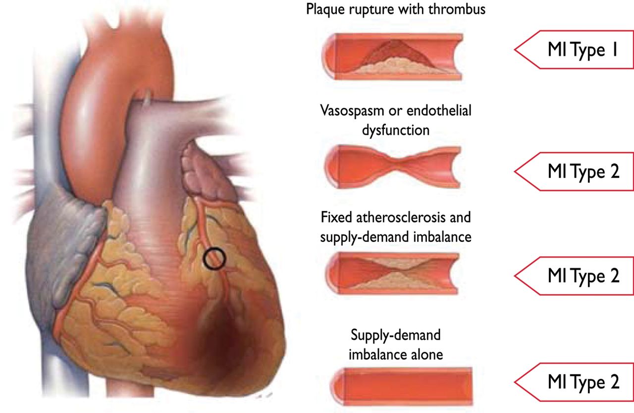
Myocardial infarction secondary to an ischaemic imbalance (MI type 2)
In instances of myocardial injury with necrosis, where a condition other than CAD contributes to an imbalance between myocardial oxygen supply and/or demand, the term ‘MI type 2’ is employed (Figure 2). In critically ill patients, or in patients undergoing major (non-cardiac) surgery, elevated values of cardiac biomarkers may appear, due to the direct toxic effects of endogenous or exogenous high circulating catecholamine levels. Also coronary vasospasm and/or endothelial dysfunction have the potential to cause MI.26–28

Differentiation between myocardial infarction (MI) types 1 and 2 according to the condition of the coronary arteries.
Cardiac death due to myocardial infarction (MI type 3)
Patients who suffer cardiac death, with symptoms suggestive of myocardial ischaemia accompanied by presumed new ischaemic ECG changes or new LBBB—but without available biomarker values—represent a challenging diagnostic group. These individuals may die before blood samples for biomarkers can be obtained, or before elevated cardiac biomarkers can be identified. If patients present with clinical features of myocardial ischaemia, or with presumed new ischaemic ECG changes, they should be classified as having had a fatal MI, even if cardiac biomarker evidence of MI is lacking.
Myocardial infarction associated with revascularization procedures (MI types 4 and 5)
Periprocedural myocardial injury or infarction may occur at some stages in the instrumentation of the heart that is required during mechanical revascularization procedures, either by PCI or by coronary artery bypass grafting (CABG). Elevated cTn values may be detected following these procedures, since various insults may occur that can lead to myocardial injury with necrosis.29–32 It is likely that limitation of such injury is beneficial to the patient: however, a threshold for a worsening prognosis, related to an asymptomatic increase of cardiac biomarker values in the absence of procedural complications, is not well defined.33–35 Subcategories of PCI-related MI are connected to stent thrombosis and restenosis that may happen after the primary procedure.
Electrocardiographic detection of myocardial infarction
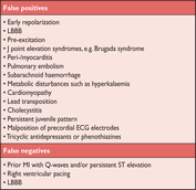
The ECG is an integral part of the diagnostic work-up of patients with suspected MI and should be acquired and interpreted promptly (i.e. target within 10 min) after clinical presentation.2 Dynamic changes in the ECG waveforms during acute myocardial ischaemic episodes often require acquisition of multiple ECGs, particularly if the ECG at initial presentation is non-diagnostic. Serial recordings in symptomatic patients with an initial non-diagnostic ECG should be performed at 15-30 min intervals or, if available, continuous computer-assisted 12-lead ECG recording. Recurrence of symptoms after an asymptomatic interval are an indication for a repeat tracing and, in patients with evolving ECG abnormalities, a pre-discharge ECG should be acquired as a baseline for future comparison. Acute or evolving changes in the ST–T waveforms and Q waves, when present, potentially allow the clinician to time the event, to identify the infarct-related artery, to estimate the amount of myocardium at risk as well as prognosis, and to determine therapeutic strategy. More profound ST-segment shift or T wave inversion involving multiple leads/territories is associated with a greater degree of myocardial ischaemia and a worse prognosis. Other ECG signs associated with acute myocardial ischaemia include cardiac arrhythmias, intraventricular and atrioventricular conduction delays, and loss of pre-cordial R wave amplitude. Coronary artery size and distribution of arterial segments, collateral vessels, location, extent and severity of coronary stenosis, and prior myocardial necrosis can all impact ECG manifestations of myocardial ischaemia.36 Therefore the ECG at presentation should always be compared to prior ECG tracings, when available. The ECG by itself is often insufficient to diagnose acute myocardial ischaemia or infarction, since ST deviation may be observed in other conditions, such as acute pericarditis, left ventricular hypertrophy (LVH), left bundle branch block (LBBB), Brugada syndrome, stress cardiomyopathy, and early repolarization patterns.37 Prolonged new ST-segment elevation (e.g. >20 min), particularly when associated with reciprocal ST-segment depression, usually reflects acute coronary occlusion and results in myocardial injury with necrosis. As in cardiomyopathy, Q waves may also occur due to myocardial fibrosis in the absence of CAD.
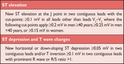
ECG abnormalities of myocardial ischaemia or infarction may be inscribed in the PR segment, the QRS complex, the ST-segment or the T wave. The earliest manifestations of myocardial ischaemia are typically T wave and ST-segment changes. Increased hyperacute T wave amplitude, with prominent symmetrical T waves in at least two contiguous leads, is an early sign that may precede the elevation of the ST-segment. Transient Q waves may be observed during an episode of acute ischaemia or (rarely) during acute MI with successful reperfusion. Table 3 lists ST–T wave criteria for the diagnosis of acute myocardial ischaemia that may or may not lead to MI. The J point is used to determine the magnitude of the ST-segment shift. New, or presumed new, J point elevation ≥0.1mV is required in all leads other than V2 and V3. In healthy men under age 40, J-point elevation can be as much as 0.25 mV in leads V2 or V3, but it decreases with increasing age. Sex differences require different cut-points for women, since J point elevation in healthy women in leads V2 and V3 is less than in men.38 ‘Contiguous leads’ refers to lead groups such as anterior leads (V1–V6), inferior leads (II, III, aVF) or lateral/apical leads (I, aVL). Supplemental leads such as V3R and V4R reflect the free wall of the right ventricle and V7–V9 the infero-basal wall.
ECG manifestations of acute myocardial ischaemia (in absence of LVH and LBBB)
 |
|
ECG manifestations of acute myocardial ischaemia (in absence of LVH and LBBB)
|
|
The criteria in Table 3 require that the ST shift be present in two or more contiguous leads. For example, ≥0.2 mV of ST elevation in lead V2, and ≥0.1 mV in lead V1, would meet the criteria of two abnormal contiguous leads in a man >40 years old. However, ≥0.1mV and <0.2mV of ST elevation, seen only in leads V2-V3 in men (or <0.15mV in women), may represent a normal finding. It should be noted that, occasionally, acute myocardial ischaemia may create sufficient ST-segment shift to meet the criteria in one lead but have slightly less than the required ST shift in a contiguous lead. Lesser degrees of ST displacement or T wave inversion do not exclude acute myocardial ischaemia or evolving MI, since a single static recording may miss the more dynamic ECG changes that might be detected with serial recordings. ST elevation or diagnostic Q waves in contiguous lead groups are more specific than ST depression in localizing the site of myocardial ischaemia or necrosis.39,40 Supplemental leads, as well as serial ECG recordings, should always be considered in patients that present with ischaemic chest pain and a non-diagnostic initial ECG.41,42 Electrocardiographic evidence of myocardial ischaemia in the distribution of a left circumflex artery is often overlooked and is best captured using posterior leads at the fifth intercostal space (V7 at the left posterior axillary line, V8 at the left mid-scapular line, and V9 at the left paraspinal border). Recording of these leads is strongly recommended in patients with high clinical suspicion for acute circumflex occlusion (for example, initial ECG non-diagnostic, or ST-segment depression in leads V1–3).41 A cut-point of 0.05 mV ST elevation is recommended in leads V7–V9; specificity is increased at a cut-point ≥0.1 mV ST elevation and this cut-point should be used in men <40 years old. ST depression in leads V1–V3 may be suggestive of infero-basal myocardial ischaemia (posterior infarction), especially when the terminal T wave is positive (ST elevation equivalent), however this is non-specific.41–43 In patients with inferior and suspected right ventricular infarction, right pre-cordial leads V3R and V4R should be recorded, since ST elevation ≥0.05 mV (≥0.1 mV in men <30 years old) provides supportive criteria for the diagnosis.42
During an episode of acute chest discomfort, pseudo-normalization of previously inverted T waves may indicate acute myocardial ischaemia. Pulmonary embolism, intracranial processes, electrolyte abnormalities, hypothermia, or peri-/myocarditis may also result in ST–T abnormalities and should be considered in the differential diagnosis. The diagnosis of MI is more difficult in the presence of LBBB.44,45 However, concordant ST-segment elevation or a previous ECG may be helpful to determine the presence of acute MI in this setting. In patients with right bundle branch block (RBBB), ST–T abnormalities in leads V1–V3 are common, making it difficult to assess the presence of ischaemia in these leads: however, when new ST elevation or Q waves are found, myocardial ischaemia or infarction should be considered.
Prior myocardial infarction
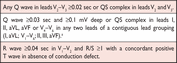
As shown in Table 4, Q waves or QS complexes in the absence of QRS confounders are pathognomonic of a prior MI in patients with ischaemic heart disease, regardless of symptoms.46,47 The specificity of the ECG diagnosis for MI is greatest when Q waves occur in several leads or lead groupings. When the Q waves are associated with ST deviations or T wave changes in the same leads, the likelihood of MI is increased; for example, minor Q waves ≥0.02 sec and <0.03 sec that are ≧ 0.1 mV deep are suggestive of prior MI if accompanied by inverted T waves in the same lead group. Other validated MI coding algorithms, such as the Minnesota Code and WHO MONICA, have been used in epidemiological studies and clinical trials.3
ECG changes associated with prior myocardial infarction
 |
|
aThe same criteria are used for supplemental leads V7–V9.
ECG changes associated with prior myocardial infarction
|
|
aThe same criteria are used for supplemental leads V7–V9.
Silent myocardial infarction
Asymptomatic patients who develop new pathologic Q wave criteria for MI detected during routine ECG follow-up, or reveal evidence of MI by cardiac imaging, that cannot be directly attributed to a coronary revascularization procedure, should be termed ‘silent MI’.48–51 In studies, silent Q wave MI accounted for 9–37% of all non-fatal MI events and were associated with a significantly increased mortality risk.48,49 Improper lead placement or QRS confounders may result in what appear to be new Q waves or QS complexes, as compared to a prior tracing. Thus, the diagnosis of a new silent Q wave MI should be confirmed by a repeat ECG with correct lead placement, or by an imaging study, and by focussed questioning about potential interim ischaemic symptoms.
Conditions that confound the ECG diagnosis of myocardial infarction
A QS complex in lead V1 is normal. A Q wave <0.03 sec and <25% of the R wave amplitude in lead III is normal if the frontal QRS axis is between -30° and 0°. A Q wave may also be normal in aVL if the frontal QRS axis is between 60° and 90°. Septal Q waves are small, non-pathological Q waves <0.03 sec and <25% of the R-wave amplitude in leads I, aVL, aVF, and V4–V6. Pre-excitation, obstructive, dilated or stress cardiomyopathy, cardiac amyloidosis, LBBB, left anterior hemiblock, LVH, right ventricular hypertrophy, myocarditis, acute cor pulmonale, or hyperkalaemia may be associated with Q waves or QS complexes in the absence of MI. ECG abnormalities that mimic myocardial ischaemia or MI are presented in Table 5.
Imaging techniques
Non-invasive imaging plays many roles in patients with known or suspected MI, but this section concerns only its role in the diagnosis and characterisation of MI. The underlying rationale is that regional myocardial hypoperfusion and ischaemia lead to a cascade of events, including myocardial dysfunction, cell death and healing by fibrosis. Important imaging parameters are therefore perfusion, myocyte viability, myocardial thickness, thickening and motion, and the effects of fibrosis on the kinetics of paramagnetic or radio-opaque contrast agents.
Commonly used imaging techniques in acute and chronic infarction are echocardiography, radionuclide ventriculography, myocardial perfusion scintigraphy (MPS) using single photon emission computed tomography (SPECT), and magnetic resonance imaging (MRI). Positron emission tomography (PET) and X-ray computed tomography (CT) are less common.52 There is considerable overlap in their capabilities and each of the techniques can, to a greater or lesser extent, assess myocardial viability, perfusion, and function. Only the radionuclide techniques provide a direct assessment of myocyte viability, because of the inherent properties of the tracers used. Other techniques provide indirect assessments of myocardial viability, such as contractile response to dobutamine by echocardiography or myocardial fibrosis by MR.
Echocardiography
The strength of echocardiography is the assessment of cardiac structure and function, in particular myocardial thickness, thickening and motion. Echocardiographic contrast agents can improve visualisation of the endocardial border and can be used to assess myocardial perfusion and microvascular obstruction. Tissue Doppler and strain imaging permit quantification of global and regional function.53 Intravascular echocardiographic contrast agents have been developed that target specific molecular processes, but these techniques have not yet been applied in the setting of MI.54
Radionuclide imaging
Several radionuclide tracers allow viable myocytes to be imaged directly, including the SPECT tracers thallium-201, technetium-99m MIBI and tetrofosmin, and the PET tracers F-2-fluorodeoxyglucose (FDG) and rubidium-82.18,52 The strength of the SPECT techniques is that these are the only commonly available direct methods of assessing viability, although the relatively low resolution of the images leaves them at a disadvantage for detecting small areas of MI. The common SPECT radiopharmaceuticals are also tracers of myocardial perfusion and the techniques thereby readily detect areas of MI and inducible perfusion abnormalities. ECG-gated imaging provides a reliable assessment of myocardial motion, thickening and global function. Evolving radionuclide techniques that are relevant to the assessment of MI include imaging of sympathetic innervation using iodine-123-labelled meta-iodo-benzylguanidine (mIBG),55 imaging of matrix metalloproteinase activation in ventricular remodelling,56,57 and refined assessment of myocardial metabolism.58
Magnetic resonance imaging
The high tissue contrast of cardiovascular MRI provides an accurate assessment of myocardial function and it has similar capability to echocardiography in suspected acute MI. Paramagnetic contrast agents can be used to assess myocardial perfusion and the increase in extracellular space that is associated with the fibrosis of prior MI. These techniques have been used in the setting of acute MI,59,60 and imaging of myocardial fibrosis by delayed contrast enhancement is able to detect even small areas of subendocardial MI. It is also of value in detecting myocardial disease states that can mimic MI, such as myocarditis.61.
Computed tomography
Infarcted myocardium is initially visible as a focal area of decreased left ventricle (LV) enhancement, but later imaging shows hyper-enhancement, as with late gadolinium imaging by MRI.62 This finding is clinically relevant because contrast-enhanced CT may be performed for suspected pulmonary embolism and aortic dissection—conditions with clinical features that overlap with those of acute MI—but the technique is not used routinely. Similarly, CT assessment of myocardial perfusion is technically feasible but not yet fully validated.
Applying imaging in acute myocardial infarction
Imaging techniques can be useful in the diagnosis of acute MI because of their ability to detect wall motion abnormalities or loss of viable myocardium in the presence of elevated cardiac biomarker values. If, for some reason, biomarkers have not been measured or may have normalized, demonstration of new loss of myocardial viability in the absence of non-ischaemic causes meets the criteria for MI. Normal function and viability have a very high negative predictive value and practically exclude acute MI.63 Thus, imaging techniques are useful for early triage and discharge of patients with suspected MI. However, if biomarkers have been measured at appropriate times and are normal, this excludes an acute MI and takes precedence over the imaging criteria.
Abnormal regional myocardial motion and thickening may be caused by acute MI or by one or more of several other conditions, including prior MI, acute ischaemia, stunning or hibernation. Non-ischaemic conditions, such as cardiomyopathy and inflammatory or infiltrative diseases, can also lead to regional loss of viable myocardium or functional abnormality. Therefore, the positive predictive value of imaging for acute MI is not high unless these conditions can be excluded, and unless a new abnormality is detected or can be presumed to have arisen in the setting of other features of acute MI.
Echocardiography provides an assessment of many non-ischaemic causes of acute chest pain, such as peri-myocarditis, valvular heart disease, cardiomyopathy, pulmonary embolism or aortic dissection.53 It is the imaging technique of choice for detecting complications of acute MI, including myocardial free wall rupture, acute ventricular septal defect, and mitral regurgitation secondary to papillary muscle rupture or ischaemia.
Radionuclide imaging can be used to assess the amount of myocardium that is salvaged by acute revascularization.64 Tracer is injected at the time of presentation, with imaging deferred until after revascularization, providing a measure of myocardium at risk. Before discharge, a second resting injection provides a measure of final infarct size, and the difference between the two corresponds to the myocardium that has been salvaged.
Applying imaging in late presentation of myocardial infarction
In case of late presentation after suspected MI, the presence of regional wall motion abnormality, thinning or scar in the absence of non-ischaemic causes, provides evidence of past MI. The high resolution and specificity of late gadolinium enhancement MRI for the detection of myocardial fibrosis has made this a very valuable technique. In particular, the ability to distinguish between subendocardial and other patterns of fibrosis provides a differentiation between ischaemic heart disease and other myocardial abnormalities. Imaging techniques are also useful for risk stratification after a definitive diagnosis of MI. The detection of residual or remote ischaemia and/or ventricular dysfunction provides powerful indicators of later outcome.
Diagnostic criteria for myocardial infarction with PCI (MI type 4)
Balloon inflation during PCI often causes transient ischaemia, whether or not it is accompanied by chest pain or ST–T changes. Myocardial injury with necrosis may result from recognizable peri-procedural events—alone or in combination—such as coronary dissection, occlusion of a major coronary artery or a side-branch, disruption of collateral flow, slow flow or no-reflow, distal embolization, and microvascular plugging. Embolization of intracoronary thrombus or atherosclerotic particulate debris may not be preventable, despite current anticoagulant and antiplatelet adjunctive therapy, aspiration or protection devices. Such events induce inflammation of the myocardium surrounding islets of myocardial necrosis.65 New areas of myocardial necrosis have been demonstrated by MRI following PCI.66
The occurrence of procedure-related myocardial cell injury with necrosis can be detected by measurement of cardiac biomarkers before the procedure, repeated 3–6 h later and, optionally, further re-measurement 12 h thereafter. Increasing levels can only be interpreted as procedure-related myocardial injury if the pre-procedural cTn value is normal (≤99th percentile URL) or if levels are stable or falling.67,68 In patients with normal pre-procedural values, elevation of cardiac biomarker values above the 99th percentile URL following PCI are indicative of procedure-related myocardial injury. In earlier studies, increased values of post-procedural cardiac biomarkers, especially CKMB, were associated with impaired outcome.69,70 However, when cTn concentrations are normal before PCI and become abnormal after the procedure, the threshold above the 99th percentile URL—whereby an adverse prognosis is evident—is not well defined71 and it is debatable whether such a threshold even exists.72 If a single baseline cTn value is elevated, it is impossible to determine whether further increases are due to the procedure or to the initial process causing the elevation. In this situation, it appears that the prognosis is largely determined by the pre-procedural cTn level.71 These relationships will probably become even more complex for the new high-sensitivity troponin assays.70
In patients undergoing PCI with normal (≤99th percentile URL) baseline cTn concentrations, elevations of cTn >5 x 99th percentile URL occurring within 48 h of the procedure—plus either (i) evidence of prolonged ischaemia (≥20 min) as demonstrated by prolonged chest pain, or (ii) ischaemic ST changes or new pathological Q waves, or (iii) angiographic evidence of a flow limiting complication, such as of loss of patency of a side branch, persistent slow-flow or no-reflow, embolization, or (iv) imaging evidence of new loss of viable myocardium or new regional wall motion abnormality—is defined as PCI-related MI (type 4a). This threshold of cTn values >5 x 99th percentile URL is arbitrarily chosen, based on clinical judgement and societal implications of the label of peri-procedural MI. When a cTn value is ≤5 x 99th percentile URL after PCI and the cTn value was normal before the PCI—or when the cTn value is >5 x 99th percentile URL in the absence of ischaemic, angiographic or imaging findings—the term ‘myocardial injury’ should be used.
If the baseline cTn values are elevated and are stable or falling, then a rise of >20% is required for the diagnosis of a type 4a MI, as with reinfarction. Recent data suggest that, when PCI is delayed after MI until biomarker concentrations are falling or have normalized, and elevation of cardiac biomarker values then reoccurs, this may have some long-term significance. However, additional data are needed to confirm this finding.73
A subcategory of PCI-related MI is stent thrombosis, as documented by angiography and/or at autopsy and a rise and/or fall of cTn values >99th percentile URL (identified as MI type 4b). In order to stratify the occurrence of stent thrombosis in relation to the timing of the PCI procedure, the Academic Research Consortium recommends temporal categories of ‘early’ (0 to 30 days), ‘late’ (31 days to 1 year), and ‘very late’ (>1 year) to distinguish likely differences in the contribution of the various pathophysiological processes during each of these intervals.74 Occasionally, MI occurs in the clinical setting of what appears to be a stent thrombosis: however, at angiography, restenosis is observed without evidence of thrombus (see section on clinical trials).
Diagnostic criteria for myocardial infarction with CABG (MI type 5)
During CABG, numerous factors can lead to periprocedural myocardial injury with necrosis. These include direct myocardial trauma from (i) suture placement or manipulation of the heart, (ii) coronary dissection, (iii) global or regional ischaemia related to inadequate intra-operative cardiac protection, (iv) microvascular events related to reperfusion, (v) myocardial injury induced by oxygen free radical generation, or (vi) failure to reperfuse areas of the myocardium that are not subtended by graftable vessels.75–77 MRI studies suggest that most necrosis in this setting is not focal but diffuse and localized in the subendocardium.78
In patients with normal values before surgery, any increase of cardiac biomarker values after CABG indicates myocardial necrosis, implying that an increasing magnitude of biomarker concentrations is likely to be related to an impaired outcome. This has been demonstrated in clinical studies employing CKMB, where elevations 5, 10 and 20 times the URL after CABG were associated with worsened prognosis; similarly, impaired outcome has been reported when cTn values were elevated to the highest quartile or quintile of the measurements.79–83
Unlike prognosis, scant literature exists concerning the use of biomarkers for defining an MI related to a primary vascular event in a graft or native vessel in the setting of CABG. In addition, when the baseline cTn value is elevated (>99th percentile URL), higher levels of biomarker values are seen post-CABG. Therefore, biomarkers cannot stand alone in diagnosing MI in this setting. In view of the adverse impact on survival observed in patients with significant elevation of biomarker concentrations, this Task Force suggests, by arbitrary convention, that cTn values >10 x 99th percentile URL during the first 48 h following CABG, occurring from a normal baseline cTn value (≤99th percentile URL). In addition, either (i) new pathological Q waves or new LBBB, or (ii) angiographically documented new graft or new native coronary artery occlusion, or (iii) imaging evidence of new loss of viable myocardium or new regional wall motion abnormality, should be considered as diagnostic of a CABG-related MI (type 5). Cardiac biomarker release is considerably higher after valve replacement with CABG than with bypass surgery alone, and with on-pump CABG compared to off-pump CABG.84 The threshold described above is more robust for isolated on-pump CABG. As for PCI, the existing principles from the universal definition of MI should be applied for the definition of MI >48 h after surgery.
Assessment of MI in patients undergoing other cardiac procedures
New ST–T abnormalities are common in patients who undergo cardiac surgery. When new pathological Q waves appear in different territories than those identified before surgery, MI (types 1 or 2) should be considered, particularly if associated with elevated cardiac biomarker values, new wall motion abnormalities or haemodynamic instability.
Novel procedures such as transcatheter aortic valve implantation (TAVI) or mitral clip may cause myocardial injury with necrosis, both by direct trauma to the myocardium and by creating regional ischaemia from coronary obstruction or embolization. It is likely that, similarly to CABG, the more marked the elevation of the biomarker values, the worse the prognosis—but data on that are not available.
Modified criteria have been proposed for the diagnosis of periprocedural MI ≤72 h after aortic valve implantation.85 However, given that there is too little evidence, it appears reasonable to apply the same criteria for procedure-related MI as stated above for CABG.
Ablation of arrhythmias involves controlled myocardial injury with necrosis, by application of warming or cooling of the tissue. The extent of the injury with necrosis can be assessed by cTn measurement: however, an elevation of cTn values in this context should not be labelled as MI.
Myocardial infarction associated with non-cardiac procedures
Perioperative MI is the most common major perioperative vascular complication in major non-cardiac surgery, and is associated with a poor prognosis.86,87 Most patients who have a perioperative MI will not experience ischaemic symptoms. Nevertheless, asymptomatic perioperative MI is as strongly associated with 30-day mortality, as is symptomatic MI.86 Routine monitoring of cardiac biomarkers in high-risk patients, both prior to and 48–72 h after major surgery, is therefore recommended. Measurement of high-sensitivity cTn in post-operative samples reveals that 45% of patients have levels above the 99th percentile URL and 22% have an elevation and a rising pattern of values indicative of evolving myocardial necrosis.88 Studies of patients undergoing major non-cardiac surgery strongly support the idea that many of the infarctions diagnosed in this context are caused by a prolonged imbalance between myocardial oxygen supply and demand, against a background of CAD.89,90 Together with a rise and/or fall of cTn values, this indicates MI type 2. However, one pathological study of fatal perioperative MI patients showed plaque rupture and platelet aggregation, leading to thrombus formation, in approximately half of such events;91 that is to say, MI type 1. Given the differences that probably exist in the therapeutic approaches to each, close clinical scrutiny and judgement is needed.
Myocardial infarction in the intensive care unit
Elevations of cTn values are common in patients in the intensive care unit and are associated with adverse prognosis, regardless of the underlying disease state.92,93 Some elevations may reflect MI type 2 due to underlying CAD and increased myocardial oxygen demand.94 Other patients may have elevated values of cardiac biomarkers, due to myocardial injury with necrosis induced by catecholamine or direct toxic effect from circulating toxins. Moreover, in some patients, MI type 1 may occur. It is often a challenge for the clinician, caring for a critically ill patient with severe single organ or multi-organ pathology, to decide on a plan of action when the patient has elevated cTn values. If and when the patient recovers from the critical illness, clinical judgement should be employed to decide whether—and to what extent—further evaluation for CAD or structural heart disease is indicated.95
Recurrent myocardial infarction
‘Incident MI’ is defined as the individual's first MI. When features of MI occur in the first 28 days after an incident event, this is not counted as a new event for epidemiological purposes. If characteristics of MI occur after 28 days following an incident MI, it is considered to be a recurrent MI.3
Reinfarction
The term ‘reinfarction’ is used for an acute MI that occurs within 28 days of an incident- or recurrent MI.3 The ECG diagnosis of suspected reinfarction following the initial MI may be confounded by the initial evolutionary ECG changes. Reinfarction should be considered when ST elevation ≥0.1 mV recurs, or new pathognomonic Q waves appear, in at least two contiguous leads, particularly when associated with ischaemic symptoms for 20 min or longer. Re-elevation of the ST-segment can, however, also be seen in threatened myocardial rupture and should lead to additional diagnostic workup. ST depression or LBBB alone are non-specific findings and should not be used to diagnose reinfarction.
In patients in whom reinfarction is suspected from clinical signs or symptoms following the initial MI, an immediate measurement of cTn is recommended. A second sample should be obtained 3–6 h later. If the cTn concentration is elevated, but stable or decreasing at the time of suspected reinfarction, the diagnosis of reinfarction requires a 20% or greater increase of the cTn value in the second sample. If the initial cTn concentration is normal, the criteria for new acute MI apply.
Myocardial injury or infarction associated with heart failure
Depending on the assay used, detectable-to-clearly elevated cTn values, indicative of myocardial injury with necrosis, may be seen in patients with HF syndrome.96 Using high-sensitivity cTn assays, measurable cTn concentrations may be present in nearly all patients with HF, with a significant percentage exceeding the 99th percentile URL, particularly in those with more severe HF syndrome, such as in acutely decompensated HF.97
Whilst MI type 1 is an important cause of acutely decompensated HF—and should always be considered in the context of an acute presentation—elevated cTn values alone, in a patient with HF syndrome, do not establish the diagnosis of MI type 1 and may, indeed, be seen in those with non-ischaemic HF. Beyond MI type 1, multiple mechanisms have been invoked to explain measurable-to-pathologically elevated cTn concentrations in patients with HF.96,97 For example, MI type 2 may result from increased transmural pressure, small-vessel coronary obstruction, endothelial dysfunction, anaemia or hypotension. Besides MI type 1 or 2, cardiomyocyte apoptosis and autophagy due to wall stretch has been experimentally demonstrated. Direct cellular toxicity related to inflammation, circulating neurohormones, infiltrative processes, as well as myocarditis and stress cardiomyopathy, may present with HF and abnormal cTn measurement.97
Whilst prevalent and complicating the diagnosis of MI, the presence, magnitude and persistence of cTn elevation in HF is increasingly accepted to be an independent predictor of adverse outcomes in both acute and chronic HF syndrome, irrespective of mechanism, and should not be discarded as ‘false positive’.97,98
In the context of an acutely decompensated HF presentation, cTn I or T should always be promptly measured and ECG recorded, with the goal of identifying or excluding MI type 1 as the precipitant. In this setting, elevated cTn values should be interpreted with a high level of suspicion for MI type 1 if a significant rise and/or fall of the marker are seen, or if it is accompanied by ischaemic symptoms, new ischaemic ECG changes or loss of myocardial function on non-invasive testing. Coronary artery anatomy may often be well-known; such knowledge may be used to interpret abnormal troponin results. If normal coronary arteries are present, either a type 2 MI or a non-coronary mechanism for troponin release may be invoked.97
On the other hand, when coronary anatomy is not established, the recognition of a cTn value in excess of the 99th percentile URL alone is not sufficient to make a diagnosis of acute MI due to CAD, nor is it able to identify the mechanism for the abnormal cTn value. In this setting, further information, such as myocardial perfusion studies, coronary angiography, or MRI is often required to better understand the cause of the abnormal cTn measurement. However, it may be difficult to establish the reason for cTn abnormalities, even after such investigations.96,97
Application of MI in clinical trials and quality assurance programmes
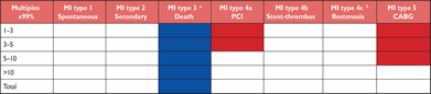
In clinical trials, MI may be an entry criterion or an end-point. A universal definition for MI is of great benefit for clinical studies, since it will allow a standardized approach for interpretation and comparison across different trials. The definition of MI as an entry criterion, e.g. MI type 1 and not MI type 2, will determine patient characteristics in the trial. Occasionally MI occurs and, at angiography, restenosis is the only angiographic explanation.99,100 This PCI-related MI type might be designated as an ‘MI type 4c’, defined as ≥50% stenosis at coronary angiography or a complex lesion associated with a rise and/or fall of cTn values >99th percentile URL and no other significant obstructive CAD of greater severity following: (i) initially successful stent deployment or (ii) dilatation of a coronary artery stenosis with balloon angioplasty (<50%).
In recent investigations, different MI definitions have been employed as trial outcomes, thereby hampering comparison and generalization between these trials. Consistency among investigators and regulatory authorities, with regard to the definition of MI used as an endpoint in clinical investigations, is of substantial value. Adaptation of the definition to an individual clinical study may be appropriate in some circumstances and should have a well-articulated rationale. No matter what, investigators should ensure that a trial provides comprehensive data for the various types of MI and includes the 99th percentile URL decision limits of cTn or other biomarkers employed. Multiples of the 99th percentiles URL may be indicated as shown in Table 6. This will facilitate comparison of trials and meta-analyses.
Tabulation in clinical trials of MI types according to multiples of the 99th percentile upper reference limit of the applied cardiac biomarker
 |
|
MI = myocardial infarction; PCI = percutaneous coronary intervention; CABG = coronary artery bypass grafting.
aBiomarker values are unavailable because of death before blood samples are obtained (blue area).
Red areas indicate arbitrarily defined cTn values below the MI decision limit whether PCI or CABG.
bRestenosis is defined as ≥50% stenosis at coronary angiography or a complex lesion associated with a rise and/or fall of cTn values >99th percentile URL and no other significant obstructive CAD of greater severity following: (i) initially successful stent deployment or (ii) dilatation of a coronary artery stenosis with balloon angioplasty (<50%).
Tabulation in clinical trials of MI types according to multiples of the 99th percentile upper reference limit of the applied cardiac biomarker
|
|
MI = myocardial infarction; PCI = percutaneous coronary intervention; CABG = coronary artery bypass grafting.
aBiomarker values are unavailable because of death before blood samples are obtained (blue area).
Red areas indicate arbitrarily defined cTn values below the MI decision limit whether PCI or CABG.
bRestenosis is defined as ≥50% stenosis at coronary angiography or a complex lesion associated with a rise and/or fall of cTn values >99th percentile URL and no other significant obstructive CAD of greater severity following: (i) initially successful stent deployment or (ii) dilatation of a coronary artery stenosis with balloon angioplasty (<50%).
Because different assays may be used, including newer, higher-sensitivity cTn assays in large multicentre clinical trials, it is advisable to consistently apply the 99th percentile URL. This will not totally harmonize troponin values across different assays, but will improve the consistency of the results. In patients undergoing cardiac procedures, the incidence of MI may be used as a measure of quality, provided that a consistent definition is applied by all centres participating in the quality assurance programme. To be effective and to avoid bias, this type of assessment will need to develop a paradigm to harmonize the different cTn assay results across sites.
Public policy implications of the adjustment of the MI definition
Revision of the definition of MI has a number of implications for individuals as well as for society at large. A tentative or final diagnosis is the basis for advice about further diagnostic testing, lifestyle changes, treatment and prognosis for the patient. The aggregate of patients with a particular diagnosis is the basis for health care planning and policy and resource allocation.
One of the goals of good clinical practice is to reach a definitive and specific diagnosis, which is supported by current scientific knowledge. The approach to the definition of MI outlined in this document meets this goal. In general, the conceptual meaning of the term ‘myocardial infarction’ has not changed, although new, sensitive diagnostic methods have been developed to diagnose this entity. Thus, the diagnosis of acute MI is a clinical diagnosis based on patient symptoms, ECG changes, and highly sensitive biochemical markers, as well as information gleaned from various imaging techniques. It is important to characterize the type of MI as well as the extent of the infarct, residual LV function, and the severity of CAD and other risk factors, rather than merely making a diagnosis of MI. The information conveyed about the patient's prognosis and ability to work requires more than just the mere statement that the patient has suffered an MI. The many additional factors just mentioned are also required so that appropriate social, family, and employment decisions can be made. A number of risk scores have been developed to predict the prognosis after MI. The classification of the various other prognostic entities associated with MI should lead to a reconsideration of the clinical coding entities currently employed for patients with the myriad conditions that can lead to myocardial necrosis, with consequent elevation of biomarker values.
It should be appreciated that the current modification of the definition of MI may be associated with consequences for the patients and their families in respect of psychological status, life insurance, professional career, as well as driving- and pilots' licences. The diagnosis is associated also with societal implications as to diagnosis-related coding, hospital reimbursement, public health statistics, sick leave, and disability attestation. In order to meet this challenge, physicians must be adequately informed of the altered diagnostic criteria. Educational materials will need to be created and treatment guidelines must be appropriately adapted. Professional societies and healthcare planners should take steps to facilitate the rapid dissemination of the revised definition to physicians, other health care professionals, administrators, and the general public.
Global perspectives of the definition of myocardial infarction
Cardiovascular disease is a global health problem. Understanding the burden and effects of CAD in populations is of critical importance. Changing clinical definitions, criteria and biomarkers add challenges to our understanding and ability to improve the health of the public. The definition of MI for clinicians has important and immediate therapeutic implications. For epidemiologists, the data are usually retrospective, so consistent case definitions are critical for comparisons and trend analysis. The standards described in this report are suitable for epidemiology studies. However, to analyse trends over time, it is important to have consistent definitions and to quantify adjustments when biomarkers or other diagnostic criteria change.101 For example, the advent of cTn dramatically increased the number of diagnosable MIs for epidemiologists.3,102
In countries with limited economic resources, cardiac biomarkers and imaging techniques may not be available except in a few centres, and even the option of ECG recordings may be lacking. In these surroundings, the WHO states that biomarker tests or other high-cost diagnostic testing are unfit for use as compulsory diagnostic criteria.3 The WHO recommends the use of the ESC/ACCF/AHA/WHF Universal MI Definition in settings without resource constraints, but recommends more flexible standards in resource-constrained locations.3
Cultural, financial, structural and organisational problems in the different countries of the world in the diagnosis and therapy of acute MI will require ongoing investigation. It is essential that the gap between therapeutic and diagnostic advances be addressed in this expanding area of cardiovascular disease.
Conflicts of interest
The members of the Task Force of the ESC, the ACCF, the AHA and the WHF have participated independently in the preparation of this document, drawing on their academic and clinical experience and applying an objective and clinical examination of all available literature. Most have undertaken—and are undertaking—work in collaboration with industry and governmental or private health providers (research studies, teaching conferences, consultation), but all believe such activities have not influenced their judgement. The best guarantee of their independence is in the quality of their past and current scientific work. However, to ensure openness, their relationships with industry, government and private health providers are reported on the ESC website (www.escardio.org/guidelines). Expenses for the Task Force/Writing Committee and preparation of this document were provided entirely by the above-mentioned joint associations.
Acknowledgements
We are very grateful to the dedicated staff of the Practice Guidelines Department of the ESC.

