


Bioresorbable stents (BRS) represent a disruptive technology with potential for improving long-term outcomes after percutaneous intervention.1 The principal advantage of BRS compared with conventional metallic stent technology is that the device is broken down by the body after its useful function has been served, potentially restoring normal vessel anatomy over the long term and ameliorating the risk of late adverse events in the treated arterial segment.2 Two such devices have received CE mark approval for use; both are based on polylactic acid stent backbones: the everolimus-eluting Absorb stent (Abbott Vascular, Abbott Park, IL, USA) in 2011 and the novolimus-eluting DESolve stent (Elixir Medical, Sunnyvale, CA, USA) in 2013. However, current approval processes leading to CE mark are based on conformity testing (i.e. performance as intended) and assessment of safety, and generally do not require comparative efficacy assessment against existing devices.3 In fact, BRS are generally more challenging to implant in comparison with metallic stents, and careful patient and lesion selection is crucial to ensuring good results with this technology.4 Moreover, clinical experience with these devices remains rather limited, and further comparative efficacy data are required before we can be sure of the place of these devices in day-to-day practice.
Encouraging results have been reported with the Absorb BRS compared with conventional metallic drug-eluting stents (DES) in small randomized trials enrolling patients presenting with stable or unstable angina.5,6 However, patients presenting with ST-elevation myocardial infarction (STEMI) might be particularly well suited to treatment with BRS, as they tend to be younger, and often have focal stenosis in relatively non-severely calcified vessels. Until now, data supporting use of BRS in STEMI have been restricted to single-centre registries without consistent long-term imaging follow-up.7–9 In the current issue of the journal, Sabate et al. report the first randomized trial comparing BRS with conventional DES in patients presenting with STEMI using 6 months optical coherence tomography (OCT) follow-up.10 Overall a total of 191 patients presenting with STEMI to eight participating clinical centres were randomized 1:1 to receive either BRS (Absorb, Abbott Vascular) or conventional permanent polymer everolimus-eluting stents (EES; Xience, Abbott Vascular).
The primary endpoint was a surrogate endpoint—which the authors termed a ‘healing score’—designed to evaluate stent–vessel wall interactions using OCT after 6 months. Secondary clinical endpoints included a device-oriented composite of cardiac death, target vessel myocardial infarction (MI), or target lesion revascularization (TLR) at 6 months, as well as device and procedural success, and stent thrombosis. The main findings can be summarized as follows: (i) implantation of the Absorb BRS results in non-inferior mean healing scores compared with EES at 6 months follow-up (scores in fact tended to be lower for Absorb); (ii) malapposed stent struts were more frequent in EES compared with Absorb; (iii) late lumen loss was significantly higher in Absorb compared with EES; and (iv) clinical outcomes were comparable in both treatment groups. Indeed, although the trial size is modest, the availability of comparative efficacy data from a randomized trial with these two technologies in patients with STEMI is an important step forward, and the authors should be congratulated for conducting such a trial in a timely manner. At the same time, the study has some limitations, which need to be considered when we interpret its results.
The most important limitations concern the primary endpoint of this trial. The healing score chosen by the authors is a multicomponent ordinal score composed of four different severity-weighted factors, i.e. the presence of intraluminal mass (weight factor of 4), malapposed and uncovered struts (weight factor of 3), uncovered but apposed struts (weight factor of 2), and malapposed and covered struts (weight factor of 1). Despite the appealing nature of adjudicating healing scores by OCT, the lack of prior validation studies establishing solid evidence for such a score is a significant issue. In fact, a major constraint of OCT in general has been its inability to detect appropriately key features of vascular healing on a cellular level owing mainly to limitations in spatial resolution.11 Stent strut coverage for instance has been widely used as a surrogate endpoint of vascular healing in clinical trials. However, the spatial resolution of OCT means that reliable detection of coverage can be challenging. In the absence of comprehensive validation studies, definitions employed in studies have ranged from a purely qualitative assessment of any tissue coverage of stent struts, to quantitative analyses applying threshold levels of tissue thickness detection taking limitations in resolution into account.12,13 In addition, any metric of tissue coverage as a binary variable fails to account for differences in quality and maturity of the tissue detected above the stent struts. While the tissue observed covering the struts could be adjudicated as mature neointimal tissue, owing to the relatively poor discriminative ability of OCT in this respect, it might just as easily be fibrin (Figure 1A and B).
![Optical coherence tomography (OCT) appearance of different arterial healing stages following implantation of everolimus-eluting (EES) permanent polymer metallic stents and Absorb bioabsorbable stents (BRS). (A) Note early arterial healing around a strut of an apposed everolimus-eluting Absorb BRS at 28 days in a healthy swine model. There is presence of fibrin/platelet deposition (*) and adjacent coverage by smooth muscle and endothelial cells as indicated by the blue arrowhead; this would be classified as healed by OCT when there is obviously incomplete healing by histology. (B) Delayed arterial healing is observed in an apposed Xience everolimus-eluting stent (EES) at 14 days from a human autopsy. Note, fibrin deposition (*) and absence of endothelial cells (green arrowhead). (C) Complete strut coverage by a fibrin/platelet thrombus of a malapposed Absorb BRS at 90 days in a healthy swine model. Note, fibrin/platelet thrombus above (yellow arrowhead) and below (*) the stent strut. Notably, differentiation of fibrin/platelet thrombus from intimal smooth muscle cells (#) is difficult with OCT. In such a situation, the strut may falsely be regarded as apposed. (D) Coverage of a malapposed Xience EES at 52 days from a human autopsy. Note, coverage by organizing thrombus (red arrowhead). Also, it is impossible to detect the presence of tissue underneath stent struts in metallic drug-eluting stents (DES) because of strut blooming artefact with extinction of light penetration (black arrowhead); consequently, appropriate determination of strut apposition may be challenging and prone to error. (E) Diagram illustrating a stent strut completely apposed to the medial wall in the early phase of healing where residual fibrin/platelet deposition is seen surrounding the strut and will be falsely classified as a healed stent strut. (F) Diagram illustrating a malapposed stent strut, where fibrin/platelet thrombus is observed circumferentially surrounding the stent strut. Note the presence of fibrin/platelet thrombus between the strut and the media [haematoxylin and eosin stain (magnification ×20)]. In E and F, pink colour represents fibrin/platelet thrombus, and blue dots represent sprouting smooth muscle and/or inflammatory cells within the fibrin matrix.](https://oup.silverchair-cdn.com/oup/backfile/Content_public/Journal/eurheartj/37/3/10.1093_eurheartj_ehv537/3/m_ehv53701.jpeg?Expires=1716424030&Signature=0WJ~E3ud4IIjUpXxecL6EQ3KduJUVMRjS9xmzoz8eEHL-bi2YwiK3Y5a2qXHn0Dr7d1Stv-PiXDOtSpiOo5DLjlwODo9d5v~BH1oehD5QVgXkMSIdWnDoiWY-CnF5diUI4E0sNi0se90Hc7-aGr3lsN3Fe3woEyDVuGQg8jzg5tBPOX-OqB3hXhRenbrxDeGA989IttMwOsV7rINlrBcX3CzXgFvu5HSS-5mElfQ1fLJvQHCb9o7tBSUur-NHaISCp9zsSbsI8WT6xn8w-JPlYFe~2tmV~gySJ~iOsoGhHoYyOCyEDOy2Gxe0xY22jpvF6Oia5QrpjFU3SBIpBDBJw__&Key-Pair-Id=APKAIE5G5CRDK6RD3PGA)
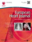
Optical coherence tomography (OCT) appearance of different arterial healing stages following implantation of everolimus-eluting (EES) permanent polymer metallic stents and Absorb bioabsorbable stents (BRS). (A) Note early arterial healing around a strut of an apposed everolimus-eluting Absorb BRS at 28 days in a healthy swine model. There is presence of fibrin/platelet deposition (*) and adjacent coverage by smooth muscle and endothelial cells as indicated by the blue arrowhead; this would be classified as healed by OCT when there is obviously incomplete healing by histology. (B) Delayed arterial healing is observed in an apposed Xience everolimus-eluting stent (EES) at 14 days from a human autopsy. Note, fibrin deposition (*) and absence of endothelial cells (green arrowhead). (C) Complete strut coverage by a fibrin/platelet thrombus of a malapposed Absorb BRS at 90 days in a healthy swine model. Note, fibrin/platelet thrombus above (yellow arrowhead) and below (*) the stent strut. Notably, differentiation of fibrin/platelet thrombus from intimal smooth muscle cells (#) is difficult with OCT. In such a situation, the strut may falsely be regarded as apposed. (D) Coverage of a malapposed Xience EES at 52 days from a human autopsy. Note, coverage by organizing thrombus (red arrowhead). Also, it is impossible to detect the presence of tissue underneath stent struts in metallic drug-eluting stents (DES) because of strut blooming artefact with extinction of light penetration (black arrowhead); consequently, appropriate determination of strut apposition may be challenging and prone to error. (E) Diagram illustrating a stent strut completely apposed to the medial wall in the early phase of healing where residual fibrin/platelet deposition is seen surrounding the strut and will be falsely classified as a healed stent strut. (F) Diagram illustrating a malapposed stent strut, where fibrin/platelet thrombus is observed circumferentially surrounding the stent strut. Note the presence of fibrin/platelet thrombus between the strut and the media [haematoxylin and eosin stain (magnification ×20)]. In E and F, pink colour represents fibrin/platelet thrombus, and blue dots represent sprouting smooth muscle and/or inflammatory cells within the fibrin matrix.
In relation to adjudication of healing scores, it must be acknowledged that because of the distinctive appearance of both stents on OCT, blinding of assessors is not feasible, and so observed differences might be due at least in part to assessor bias. In addition, the difference in healing scores in the current study was largely driven by a higher rate of malapposed stent struts in the BRS group, either covered or uncovered. However, it needs to be remembered that the determination of malapposition is fundamentally different between BRS and metallic stents due to a phenomenon called blooming artefact, which is observed when OCT signal encounters metallic surfaces causing diffuse high intensity signal artefact and extinction of signal behind the metallic surface. Histologically, malapposed stent struts are defined as struts lacking apposition to the underlying vessel wall (which may be the media or intima in the presence or absence of atherosclerosis); often fibrin-rich thrombus fills the gap between stent struts and intimal tissue during the course of healing (Figure 1C). However because of blooming artefact, fibrin thrombus underlying malapposed metallic stent struts cannot be detected, while the space between malapposed BRS struts and the intima can be readily recognized as either occupied or devoid of tissue. Moreover, because the tissue composition cannot be differentiated with OCT, malapposed BRS struts with underlying fibrin thrombus would be determined to be apposed, which is out of line with the histopathological definition of malapposition (Figure 1F).
A related unresolved issue with OCT imaging is neointimal tissue characterization after stent implantation. A number of studies have tried to address this issue. In one study by Templin et al., optical density measurements of fibrin-covered stents were compared with morphological information gathered by scanning electron microscopy (SEM).14 However, this study was limited by the inability of SEM to differentiate fibrin from other extracellular matrix compounds. We have recently studied the concept of using grey-scale signal intensity analysis of OCT images to try to distinguish mature from immature neointimal tissue after stent implantation.11 In this study, we correlated histopathological findings after stent implantation in rabbits with co-registered OCT frames. Threshold levels were established for the detection of mature neointimal tissue, and then application of these thresholds was tested in patients treated with DES undergoing routine invasive surveillance imaging follow-up, which provided evidence that the majority of tissue coverage remains immature after 6 months. In another study from Japan, six autopsy hearts with 10 bare metal stents were imaged and assessed by histopathology to investigate the tissue composition of OCT-derived homogenous and heterogenous tissue patterns with either visible or invisible struts.15 The homogenous tissue pattern mostly consisted of smooth muscle cells with collagen, indicating high neointimal maturity. The heterogenous tissue pattern with invisible struts contained a variety of different tissue components such as neointimal lipid/necrotic core, accumulation of foam cells, and/or microcalcification. In contrast, the heterogenous tissue pattern with visible stent struts was mostly composed of proteoglycan-rich myxomatous matrix or dense calcification. These studies all emphasize that appropriate histology–OCT correlation studies are required before novel healing scores can be adopted in prospective clinical trials.
From a clinical perspective, the use of the Absorb BRS in the setting of STEMI seemed feasible without major safety concerns. With accurate sizing and appropriate implantation technique, similar acute angiographic results may be obtained as compared with metallic DES. Importantly, however, the absence of any difference in clinical endpoints during follow-up should not be interpreted as evidence of absence of difference. In fact in view of the observed rate of the device-oriented composite endpoint in the study, the power of the trial to detect true differences between the two devices is very low. In addition, in view of the fact that the majority of patients presenting with STEMI to participating centres were not screened for inclusion (only 216 of a total of 2055 patients were screened), the external validity of the findings is unclear.
Finally, the clinical results of the study of Sabate et al. build on findings from the largest observational registry of BRS in STEMI published to date, which enrolled 290 patients and compared clinical outcomes of the Absorb BRS in a propensity score-matched analysis with EES and bare metal stent-treated patients.16 At 1-year follow-up in that study, the device-oriented composite endpoint of cardiac death, target vessel MI, and TLR was not different between groups, although, numerically, definite and probable stent thrombosis occurred more frequently in Absorb-treated patients. In fact, findings in relation to stent thrombosis with BRS in general have received considerable attention after publication of registry studies reporting rates higher than we have been accustomed to with current generation metallic DES,17 along with pre-clinical studies demonstrating higher thrombogenicity after BRS implantation as compared with metallic stents in the acute phase.18 Ultimately, what is required to answer questions in relation to clinical outcomes is data from large-scale comparative efficacy studies powered for hard clinical endpoints and incorporating long-term follow-up. We believe that such studies should be mandated as part of evaluation for regulatory approval in Europe in the future.3 In fact large-scale trials are ongoing, and the primary results of one trial designed in consultation with the Food and Drug Administration with a view to device approval in the USA—the ABSORB III randomized trial (clinicaltrials.gov identifier NCT01751906)—are expected to be presented in the coming weeks. Until long-term follow-up data from such trials become available, the idea of improving vessel healing—and more importantly patient outcomes—with fully bioresorbable DES remains a pipe dream.
Conflict of interest: M.J. is a consultant for Biotronik, and has received speakers fees from Abbott Vascular, Biotronik and Orbus Neich. He has received institutional grant support from Abbott Vascular, BioSensors International, Biotronik, Boston Scientific, Medtronic, MicroPort Medical, OrbusNeich Medical, SINO Medical Technology, and Terumo Corporation. T.K. reports no conflicts of interest. R.V. receives research support from Abbott Vascular, BioSensors International, Biotronik, Boston Scientific, Medtronic, MicroPort Medical, OrbusNeich Medical, SINO Medical Technology, and Terumo Corporation; has speaking engagements with Merck; receives honoraria from Abbott Vascular, Boston Scientific, Lutonix, Medtronic, and Terumo Corporation; and is a consultant for 480 Biomedical, Abbott Vascular, Medtronic, and W.L. Gore. R.A.B. has received speakers fees from B. Braun Melsungen, Biotronik, and Boston Scientific.
References
Author notes
The opinions expressed in this article are not necessarily those of the Editors of the European Heart Journal or of the European Society of Cardiology.
doi:10.1093/eurheartj/ehv500.

