


A 63-year-old female who had palpitations and dizziness was referred to our hospital because of cardiac enlargement shown on chest X-ray in a health examination. On chest X-ray, enlargement of protrusion of the right first and left first aortic arches was observed. Electrocardiogram revealed ectopic low part atrial rhythm, right axis deviation of the QRS wave, and incomplete right bundle branch block.
On transthoracic echocardiogram (TTE), atrial septal defect (ASD) with right to left shunt was observed, presence of Eisenmenger syndrome was suspected and there seemed to be no surgical indication. But on TTE, right heart configuration seemed not due to pressure load but volume load, which is not indicative of Eisenmenger syndrome.
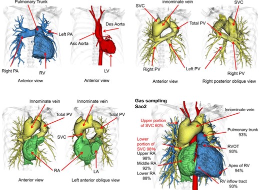
To evaluate the presence of other complications of anatomical cardiovascular heart disease, ECG-gated 320 slice CT (Aquilion ONE) was performed. On CT, supra-cardiac type total anomalous pulmonary venous return (TAPVR) with right to left shunt ASD could be accurately diagnosed. She then underwent TAPVR surgical repair from the superior approach in which total PV connected to the LA and ligated innominate vein and ASD pericardial patch closure. By this procedure, there was no blood flow from PV to the right heart, with blood from the PV flowing into the LA directly. Haemodynamics was normalized.
In summary, this patient had remarkable cardiac enlargement on chest X-ray, and on TTE ASD with right-to-left shunt was detected. 320 slice CT can evaluate complex congenital heart disease as well as coronary arteries accurately and provide information on blood flow.

